WB
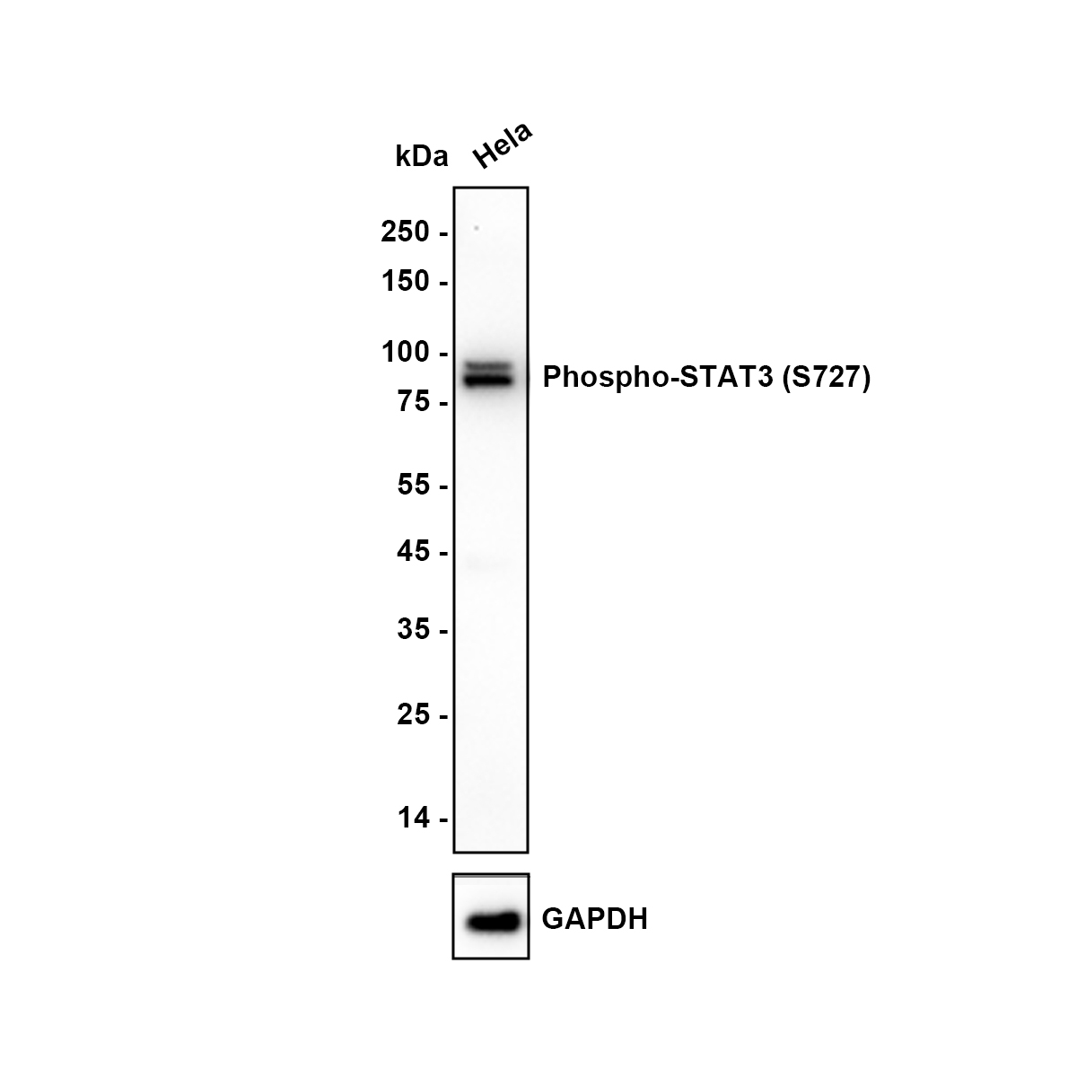
Western blot analysis of Phospho-STAT3(S727) on Hela cell lysate with Rabbit anti-Phospho-STAT3(S727) antibody at 1/1,000 dilution. Lysates/proteins at 15 µg/Lane. Exposure time: 30 seconds; Blocking: 5% NFDM/TBST, 1 hour at room temperature Primary antibody: 1/1,000 in 5% NFDM/TBST, overnight at 4 ℃ Secondary antibody: Goat anti-Rabbit IgG-HRP, 1/50,000 in 5% NFDM/TBST, 1 hour at room temperature.IHC
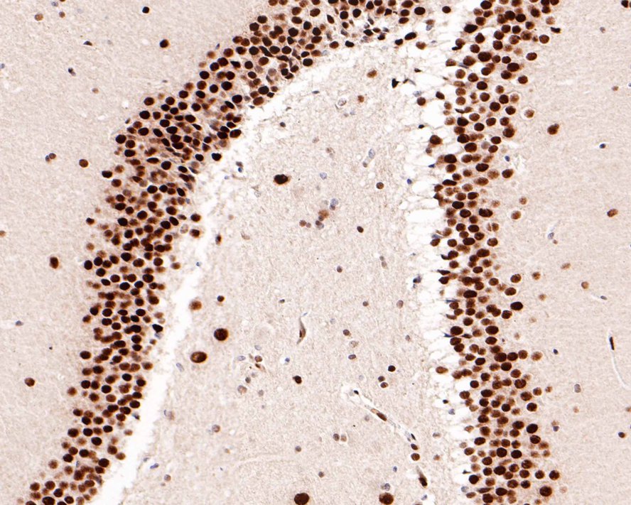
Immunohistochemical analysis of paraffin-embedded rat hippocampus tissue with Rabbit anti-Phospho-STAT3 (S727) antibody at 1:500 dilution. The section was pre-treated using heat mediated antigen retrieval with Tris-EDTA buffer (pH 9.0) for 20 minutes. The tissues were blocked in 1% BSA for 20 minutes at room temperature, washed with ddH2O and PBS, and then probed with the primary antibody at 1:500 dilution for 1 hour at room temperature. The detection was performed using an HRP conjugated compact polymer system. DAB was used as the chromogen. Tissues were counterstained with hematoxylin and mounted with DPX.IHC
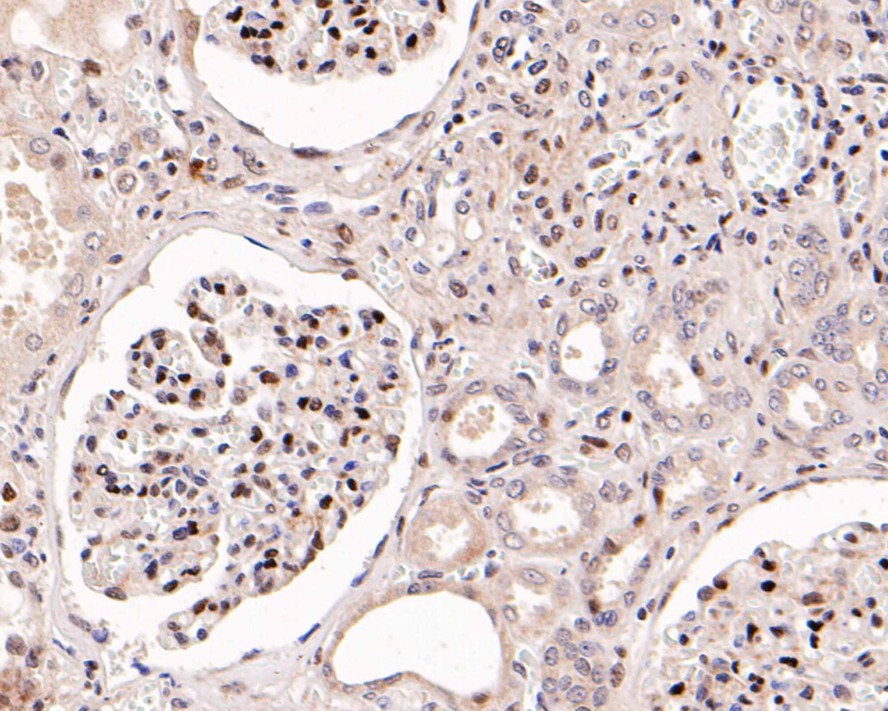
Immunohistochemical analysis of paraffin-embedded human kidney tissue with Rabbit anti-Phospho-STAT3 (S727) antibody at 1:500 dilution. The section was pre-treated using heat mediated antigen retrieval with Tris-EDTA buffer (pH 9.0) for 20 minutes. The tissues were blocked in 1% BSA for 20 minutes at room temperature, washed with ddH2O and PBS, and then probed with the primary antibody at 1:500 dilution for 1 hour at room temperature. The detection was performed using an HRP conjugated compact polymer system. DAB was used as the chromogen. Tissues were counterstained with hematoxylin and mounted with DPX.IHC
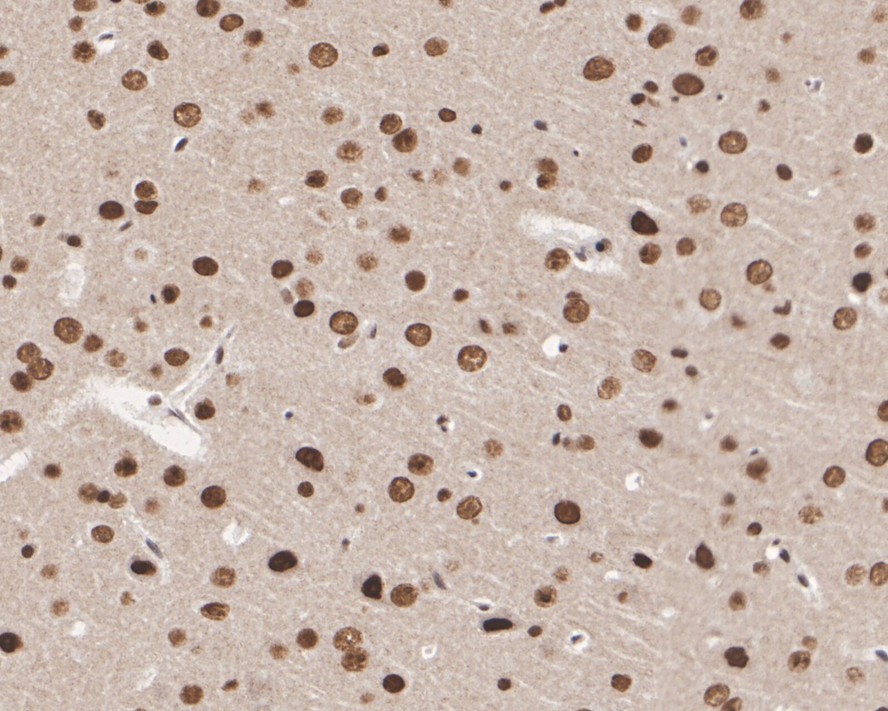
Immunohistochemical analysis of paraffin-embedded mouse brain tissue with Rabbit anti-Phospho-STAT3 (S727) antibody at 1/1,000 dilution. The section was pre-treated using heat mediated antigen retrieval with sodium citrate buffer (pH 6.0) (high pressure) for 2 minutes. The tissues were blocked in 1% BSA for 20 minutes at room temperature, washed with ddH2O and PBS, and then probed with the primary antibody at 1/1,000 dilution for 1 hour at room temperature. The detection was performed using an HRP conjugated compact polymer system. DAB was used as the chromogen. Tissues were counterstained with hematoxylin and mounted with DPX.ICC/IF
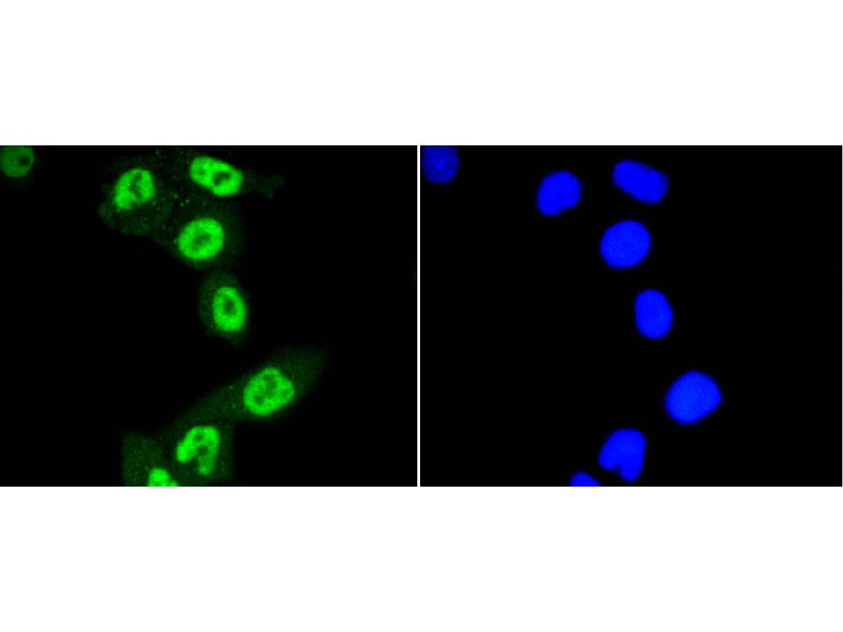
ICC staining of Phospho-STAT3 (S727) in Hela cells (green). Formalin fixed cells were permeabilized with 0.1% Triton X-100 in TBS for 10 minutes at room temperature and blocked with 1% Blocker BSA for 15 minutes at room temperature. Cells were probed with the primary antibody (1/100) for 1 hour at room temperature, washed with PBS. Alexa Fluor®488 Goat anti-Rabbit IgG was used as the secondary antibody at 1/1,000 dilution. The nuclear counter stain is DAPI (blue).ICC/IF
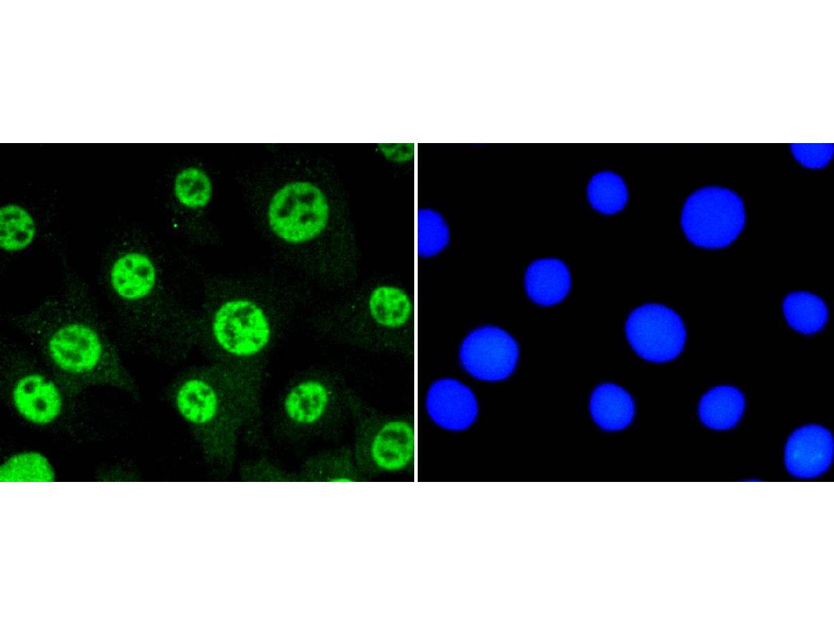
ICC staining of Phospho-STAT3 (S727) in NIH/3T3 cells (green). Formalin fixed cells were permeabilized with 0.1% Triton X-100 in TBS for 10 minutes at room temperature and blocked with 1% Blocker BSA for 15 minutes at room temperature. Cells were probed with the primary antibody (1/100) for 1 hour at room temperature, washed with PBS. Alexa Fluor®488 Goat anti-Rabbit IgG was used as the secondary antibody at 1/1,000 dilution. The nuclear counter stain is DAPI (blue).| Product Name | Phospho-STAT3 (S727) Recombinant Rabbit Monoclonal Antibody |
|---|---|
| Antibody Type | Primary Antibodies |
| Immunogen | Synthetic phospho-peptide corresponding to residues surrounding Ser727 of Human STAT3 aa 701-750 / 770. |
| Modification | p-S727 |
| Clonality | Monoclonal |
|---|---|
| Isotype | IgG |
| Host Species | Rabbit |
| Tested Applications | ICC/IFIHCWB |
| WB:1:1000-1:2000 IHC:1:500-1:1000 ICC/IF:1:100-1:200 |
|
| Species Reactivity | HumanMouseRat |
| Concentration | 1mg/ml |
| Purification | Protein A |
| Gene Symbol | STAT3 |
|---|---|
| Gene Synonyms | APRF HIES ADMIO ADMIO1 |
| Gene Full Name | signal transducer and activator of transcription 3 |
| Gene Summary | The protein encoded by this gene is a member of the STAT protein family. In response to cytokines and growth factors, STAT family members are phosphorylated by the receptor associated kinases, and then form homo- or heterodimers that translocate to the cell nucleus where they act as transcription activators. This protein is activated through phosphorylation in response to various cytokines and growth factors including IFNs, EGF, IL5, IL6, HGF, LIF and BMP2. This protein mediates the expression of a variety of genes in response to cell stimuli, and thus plays a key role in many cellular processes such as cell growth and apoptosis. The small GTPase Rac1 has been shown to bind and regulate the activity of this protein. PIAS3 protein is a specific inhibitor of this protein. This gene also plays a role in regulating host response to viral and bacterial infections. Mutations in this gene are associated with infantile-onset multisystem autoimmune disease and hyper-immunoglobulin E syndrome. [provided by RefSeq, Aug 2020] |
| Alternative Names | 1110034C02Rik antibody Acute Phase Response Factor antibody Acute-phase response factor antibody ADMIO antibody APRF antibody AW109958 antibody DNA binding protein APRF antibody FLJ20882 antibody HIES antibody MGC16063 antibody Signal transducer and activator of transcription 3 (acute phase response factor) antibody Signal transducer and activator of transcription 3 antibody STAT 3 antibody Stat3 antibody STAT3_HUMAN antibody |
| Molecular Weight(MW) | 88.1 kDa(Observed band size:88 kDa) |
| Cellular Localization | Cytoplasm, Nucleus. |
WB
Western blot analysis of Phospho-STAT3(S727) on Hela cell lysate with Rabbit anti-Phospho-STAT3(S727) antibody at 1/1,000 dilution. Lysates/proteins at 15 µg/Lane. Exposure time: 30 seconds; Blocking: 5% NFDM/TBST, 1 hour at room temperature Primary antibody: 1/1,000 in 5% NFDM/TBST, overnight at 4 ℃ Secondary antibody: Goat anti-Rabbit IgG-HRP, 1/50,000 in 5% NFDM/TBST, 1 hour at room temperature.
IHC
Immunohistochemical analysis of paraffin-embedded rat hippocampus tissue with Rabbit anti-Phospho-STAT3 (S727) antibody at 1:500 dilution. The section was pre-treated using heat mediated antigen retrieval with Tris-EDTA buffer (pH 9.0) for 20 minutes. The tissues were blocked in 1% BSA for 20 minutes at room temperature, washed with ddH2O and PBS, and then probed with the primary antibody at 1:500 dilution for 1 hour at room temperature. The detection was performed using an HRP conjugated compact polymer system. DAB was used as the chromogen. Tissues were counterstained with hematoxylin and mounted with DPX.
IHC
Immunohistochemical analysis of paraffin-embedded human kidney tissue with Rabbit anti-Phospho-STAT3 (S727) antibody at 1:500 dilution. The section was pre-treated using heat mediated antigen retrieval with Tris-EDTA buffer (pH 9.0) for 20 minutes. The tissues were blocked in 1% BSA for 20 minutes at room temperature, washed with ddH2O and PBS, and then probed with the primary antibody at 1:500 dilution for 1 hour at room temperature. The detection was performed using an HRP conjugated compact polymer system. DAB was used as the chromogen. Tissues were counterstained with hematoxylin and mounted with DPX.
IHC
Immunohistochemical analysis of paraffin-embedded mouse brain tissue with Rabbit anti-Phospho-STAT3 (S727) antibody at 1/1,000 dilution. The section was pre-treated using heat mediated antigen retrieval with sodium citrate buffer (pH 6.0) (high pressure) for 2 minutes. The tissues were blocked in 1% BSA for 20 minutes at room temperature, washed with ddH2O and PBS, and then probed with the primary antibody at 1/1,000 dilution for 1 hour at room temperature. The detection was performed using an HRP conjugated compact polymer system. DAB was used as the chromogen. Tissues were counterstained with hematoxylin and mounted with DPX.
ICC/IF
ICC staining of Phospho-STAT3 (S727) in Hela cells (green). Formalin fixed cells were permeabilized with 0.1% Triton X-100 in TBS for 10 minutes at room temperature and blocked with 1% Blocker BSA for 15 minutes at room temperature. Cells were probed with the primary antibody (1/100) for 1 hour at room temperature, washed with PBS. Alexa Fluor®488 Goat anti-Rabbit IgG was used as the secondary antibody at 1/1,000 dilution. The nuclear counter stain is DAPI (blue).
ICC/IF
ICC staining of Phospho-STAT3 (S727) in NIH/3T3 cells (green). Formalin fixed cells were permeabilized with 0.1% Triton X-100 in TBS for 10 minutes at room temperature and blocked with 1% Blocker BSA for 15 minutes at room temperature. Cells were probed with the primary antibody (1/100) for 1 hour at room temperature, washed with PBS. Alexa Fluor®488 Goat anti-Rabbit IgG was used as the secondary antibody at 1/1,000 dilution. The nuclear counter stain is DAPI (blue).| Application Notes | WB:1:1000-1:2000 IHC:1:500-1:1000 ICC/IF:1:100-1:200 |
|---|
| Form | Liquid |
|---|---|
| Storage Instructions | Shipped at 4℃. Store at +4℃ short term (1-2 weeks). Store at -20℃ long term. |
| Storage Buffer | 1*PBS (pH7.4), 0.1% BSA, 40% Glycerol, 0.2% Proclean 950. |


