WB
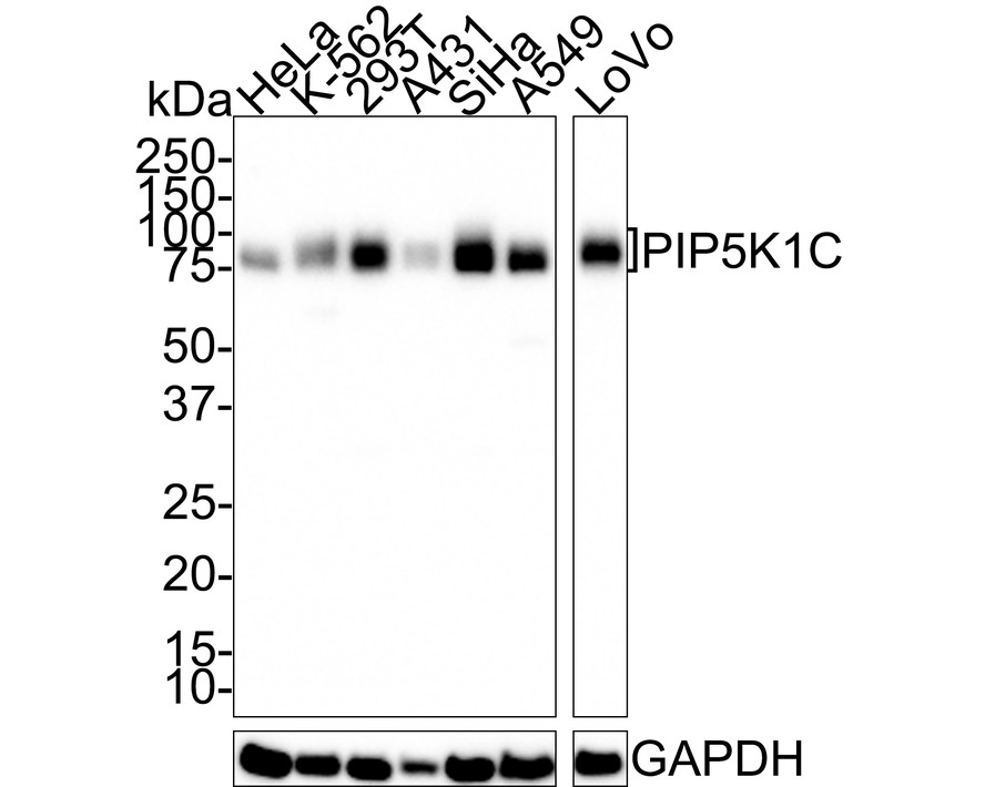
Western blot analysis of PIP5K1C on different lysates with Rabbit anti-PIP5K1C antibody at 1/1,000 dilution. Lysates/proteins at 20 µg/Lane. Exposure time: 3 minutes; 4-20% SDS-PAGE gel. Proteins were transferred to a PVDF membrane and blocked with 5% NFDM/TBST for 1 hour at room temperature. The primary antibody at 1/1,000 dilution was used in 5% NFDM/TBST at 4℃ overnight. Goat Anti-Rabbit IgG - HRP Secondary Antibody at 1/50,000 dilution was used for 1 hour at room temperature.WB
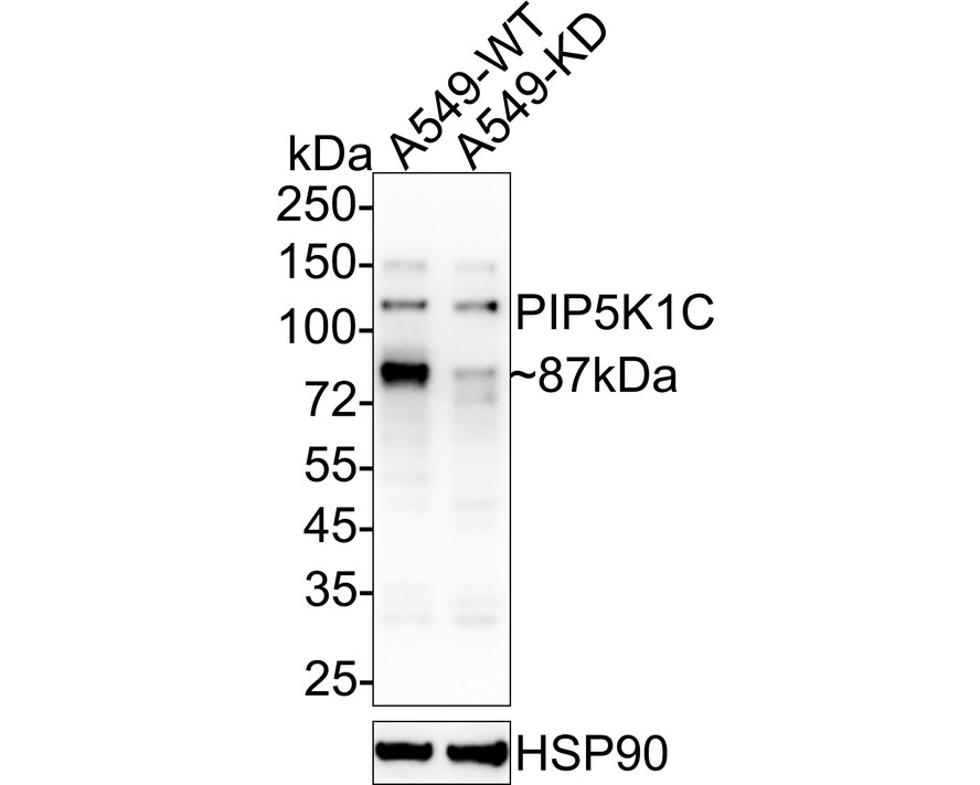
Knockdown (KD) Western blot analysis of PIP5K1C on different lysates with Rabbit anti-PIP5K1C antibody at 1/1,000 dilution. Lysates/proteins at 20 µg/Lane. Exposure time: 40 seconds; 4-20% SDS-PAGE gel. Proteins were transferred to a PVDF membrane and blocked with 5% NFDM/TBST for 1 hour at room temperature. The primary antibody at 1/1,000 dilution was used in 5% NFDM/TBST at 4℃ overnight. Goat Anti-Rabbit IgG - HRP Secondary Antibody at 1/50,000 dilution was used for 1 hour at room temperature.| Product Name | PIP5K1C Recombinant Rabbit Monoclonal Antibody |
|---|---|
| Antibody Type | Primary Antibodies |
| Immunogen | Recombinant protein within human PIP5K1C aa 31-470 / 668. |
| Clonality | Monoclonal |
|---|---|
| Isotype | IgG |
| Host Species | Rabbit |
| Tested Applications | WB |
| WB:1:1000 |
|
| Species Reactivity | Human |
| Concentration | 1mg/ml |
| Purification | Protein A |
| Gene Symbol | PIP5K1C |
|---|---|
| Gene Synonyms | LCCS3 NEDVIL PIP5Kgamma PIP5K-GAMMA PIP5K1-gamma |
| Gene Full Name | phosphatidylinositol-4-phosphate 5-kinase type 1 gamma |
| Gene Summary | This locus encodes a type I phosphatidylinositol 4-phosphate 5-kinase. The encoded protein catalyzes phosphorylation of phosphatidylinositol 4-phosphate, producing phosphatidylinositol 4,5-bisphosphate. This enzyme is found at synapses and has been found to play roles in endocytosis and cell migration. Mutations at this locus have been associated with lethal congenital contractural syndrome. Alternatively spliced transcript variants encoding different isoforms have been described.[provided by RefSeq, Sep 2010] |
| Alternative Names | Diphosphoinositide kinase antibody LCCS3 antibody Phosphatidylinositol-4-phosphate 5-kinase type I gamma antibody Phosphatidylinositol-4-phosphate 5-kinase type-1 gamma antibody PI51C antibody PI51C_HUMAN antibody PIP5K GAMMA antibody PIP5K1-gamma antibody Pip5k1c antibody PIP5KIgamma antibody PtdIns(4)P 5 kinase gamma antibody PtdIns(4)P-5-kinase 1 gamma antibody Type I PIP kinase antibody |
| Molecular Weight(MW) | 73 kDa(Observed band size:87/90 kDa) |
| Cellular Localization | Cell membrane, Endomembrane system, Cytoplasm, Cell junction, focal adhesion, adherens junction, Cell projection, ruffle membrane, phagocytic cup, uropodium; Cytoplasm, Nucleus. |
WB
Western blot analysis of PIP5K1C on different lysates with Rabbit anti-PIP5K1C antibody at 1/1,000 dilution. Lysates/proteins at 20 µg/Lane. Exposure time: 3 minutes; 4-20% SDS-PAGE gel. Proteins were transferred to a PVDF membrane and blocked with 5% NFDM/TBST for 1 hour at room temperature. The primary antibody at 1/1,000 dilution was used in 5% NFDM/TBST at 4℃ overnight. Goat Anti-Rabbit IgG - HRP Secondary Antibody at 1/50,000 dilution was used for 1 hour at room temperature.
WB
Knockdown (KD) Western blot analysis of PIP5K1C on different lysates with Rabbit anti-PIP5K1C antibody at 1/1,000 dilution. Lysates/proteins at 20 µg/Lane. Exposure time: 40 seconds; 4-20% SDS-PAGE gel. Proteins were transferred to a PVDF membrane and blocked with 5% NFDM/TBST for 1 hour at room temperature. The primary antibody at 1/1,000 dilution was used in 5% NFDM/TBST at 4℃ overnight. Goat Anti-Rabbit IgG - HRP Secondary Antibody at 1/50,000 dilution was used for 1 hour at room temperature.| Application Notes | WB:1:1000 |
|---|
| Form | Liquid |
|---|---|
| Storage Instructions | Shipped at 4℃. Store at +4℃ short term (1-2 weeks). Store at -20℃ long term. |
| Storage Buffer | PBS (pH7.4), 0.1% BSA, 40% Glycerol. Preservative: 0.05% Sodium Azide. |


