WB
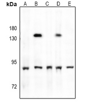
Western blot analysis of TBK1 (Phospho-S172) expression in rat testis (A), SGC7901 (B), EC9706 (C), A549 (D), 3T3L1 (E) whole cell lysates.IHC
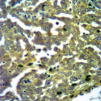
Immunohistochemical analysis of TBK1 (Phospho-S172) staining in human ovarian cancer formalin fixed paraffin embedded tissue section. The section was pre-treated using heat mediated antigen retrieval with sodium citrate buffer (pH 6.0). The section was then incubated with the antibody at room temperature and detected using an HRP conjugated compact polymer system. DAB was used as the chromogen. The section was then counterstained with haematoxylin and mounted with DPX.ICC/IF
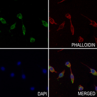
Immunofluorescent analysis of TBK1 (Phospho-S172) staining in MDAMB231 cells. Formalin-fixed cells were permeabilized with 0.1% Triton X-100 in TBS for 5-10 minutes and blocked with 3% BSA-PBS for 30 minutes at room temperature. Cells were probed with the primary antibody in 3% BSA-PBS and incubated overnight at 4 °C in a hidified chamber. Cells were washed with PBST and incubated with a AF488-conjugated secondary antibody (green) in PBS at room temperature in the dark. Phalloidin - AF594 was used to stain Actin filaments (red). DAPI was used to stain the cell nuclei (blue).| Product Name | Anti-TBK1 (Phospho-S172) Antibody |
|---|---|
| Antibody Type | Primary Antibodies |
| Immunogen | KLH-conjugated synthetic phosphopeptide corresponding to residues surrounding S172 of human TBK1 protein. The exact sequence is proprietary. |
| Modification | p-S172 |
| Clonality | Polyclonal |
|---|---|
| Isotype | IgG |
| Host Species | Rabbit |
| Tested Applications | ICC/IFIHCWB |
| WB:1:500-1:1000 IHC:1:50-1:100 ICC/IF:1:50-1:200 |
|
| Species Reactivity | HumanMouseRat |
| Concentration | 1mg/ml |
| Purification | Affinity purified |
| Gene Symbol | TBK1 |
|---|---|
| Gene Synonyms | NAK T2K AIARV IIAE8 FTDALS4 |
| Gene Full Name | TANK binding kinase 1 |
| Gene Summary | The NF-kappa-B (NFKB) complex of proteins is inhibited by I-kappa-B (IKB) proteins, which inactivate NFKB by trapping it in the cytoplasm. Phosphorylation of serine residues on the IKB proteins by IKB kinases marks them for destruction via the ubiquitination pathway, thereby allowing activation and nuclear translocation of the NFKB complex. The protein encoded by this gene is similar to IKB kinases and can mediate NFKB activation in response to certain growth factors. The protein is also an important kinase for antiviral innate immunity response. [provided by RefSeq, Sep 2021] |
| Alternative Names | NAK Serine/threonine-protein kinase TBK1 NF-kappa-B-activating kinase T2K TANK-binding kinase 1 |
| Molecular Weight(MW) | 83 kDa |
| Cellular Localization | Cytoplasm. |
WB
Western blot analysis of TBK1 (Phospho-S172) expression in rat testis (A), SGC7901 (B), EC9706 (C), A549 (D), 3T3L1 (E) whole cell lysates.
IHC
Immunohistochemical analysis of TBK1 (Phospho-S172) staining in human ovarian cancer formalin fixed paraffin embedded tissue section. The section was pre-treated using heat mediated antigen retrieval with sodium citrate buffer (pH 6.0). The section was then incubated with the antibody at room temperature and detected using an HRP conjugated compact polymer system. DAB was used as the chromogen. The section was then counterstained with haematoxylin and mounted with DPX.
ICC/IF
Immunofluorescent analysis of TBK1 (Phospho-S172) staining in MDAMB231 cells. Formalin-fixed cells were permeabilized with 0.1% Triton X-100 in TBS for 5-10 minutes and blocked with 3% BSA-PBS for 30 minutes at room temperature. Cells were probed with the primary antibody in 3% BSA-PBS and incubated overnight at 4 °C in a hidified chamber. Cells were washed with PBST and incubated with a AF488-conjugated secondary antibody (green) in PBS at room temperature in the dark. Phalloidin - AF594 was used to stain Actin filaments (red). DAPI was used to stain the cell nuclei (blue).| Application Notes | WB:1:500-1:1000 IHC:1:50-1:100 ICC/IF:1:50-1:200 |
|---|
| Form | Liquid |
|---|---|
| Storage Instructions | Shipped at 4°C. Upon delivery aliquot and store at -20°C for one year. Avoid freeze/thaw cycles. |
| Storage Buffer | Liquid in 0.42% Potassium phosphate, 0.87% Sodium chloride, pH 7.3, 30% glycerol, and 0.01% sodium azide. |


