WB
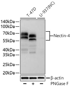
Western blot analysis of various lysates using Nectin-4 Rabbit mAb at 1:5000 dilution incubated overnight at 4℃. T-47D cells were treated with PNGase F (2 U/μL) at 37℃ for 1 hour. Secondary antibody: HRP-conjugated Goat anti-Rabbit IgG (H+L) at 1:10000 dilution. Lysates/proteins: 30 μg per lane. Blocking buffer: 3% nonfat dry milk in TBST. Detection: ECL Basic Kit. Negative control (NC): U-937. Exposure time: 90 s.IHC
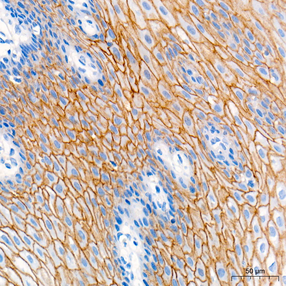
Immunohistochemistry analysis of paraffin embedded Human esophagus tissue using Nectin-4 Rabbit mAb at a dilution of 1:500 (40x lens). High pressure antigen retrieval performed with 0.01M Tris-EDTA Buffer (pH 9.0) prior to IHC staining.IHC
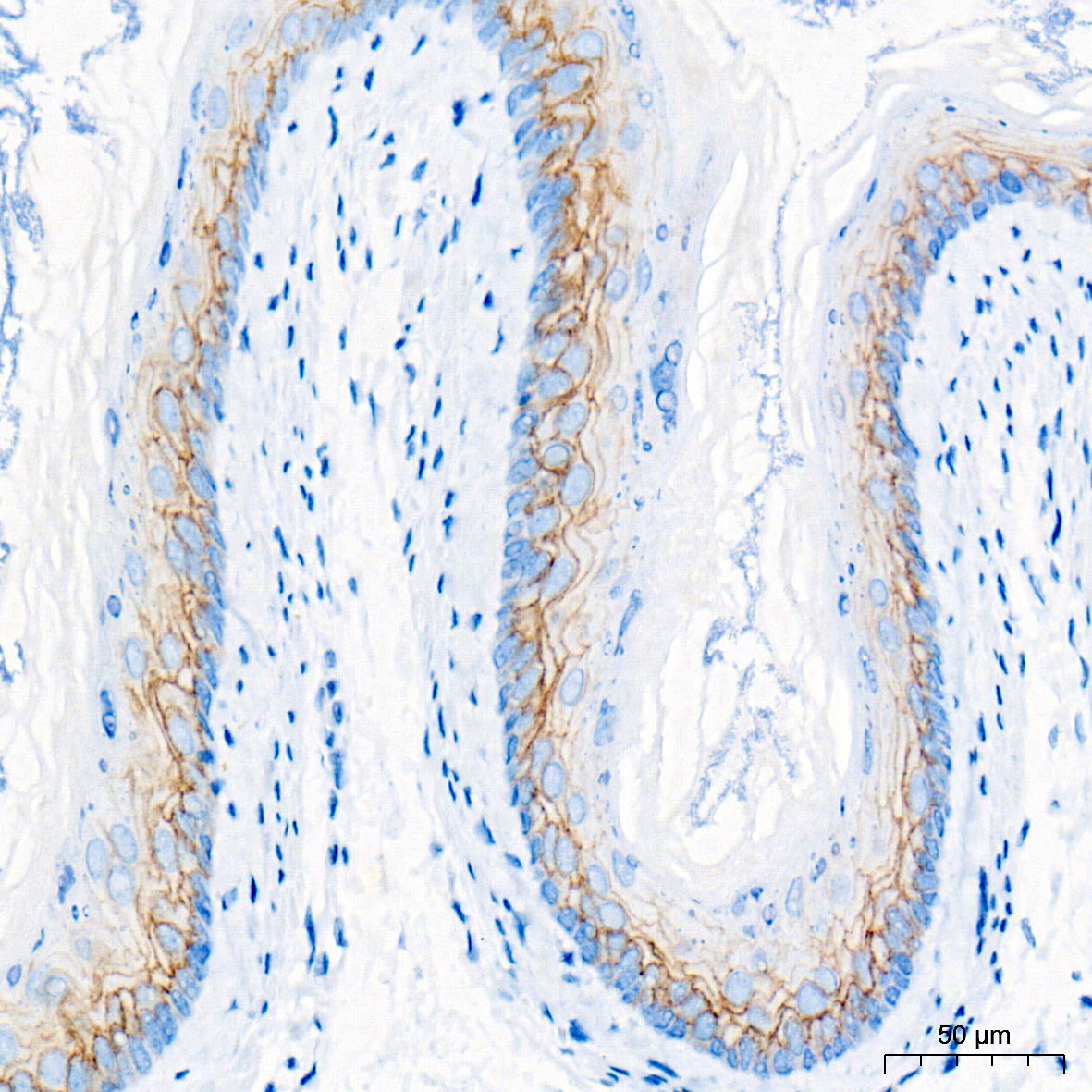
Immunohistochemistry analysis of paraffin embedded Mouse esophagus tissue using Nectin-4 Rabbit mAb at a dilution of 1:500 (40x lens). High pressure antigen retrieval performed with 0.01M Tris-EDTA Buffer (pH 9.0) prior to IHC staining.IHC
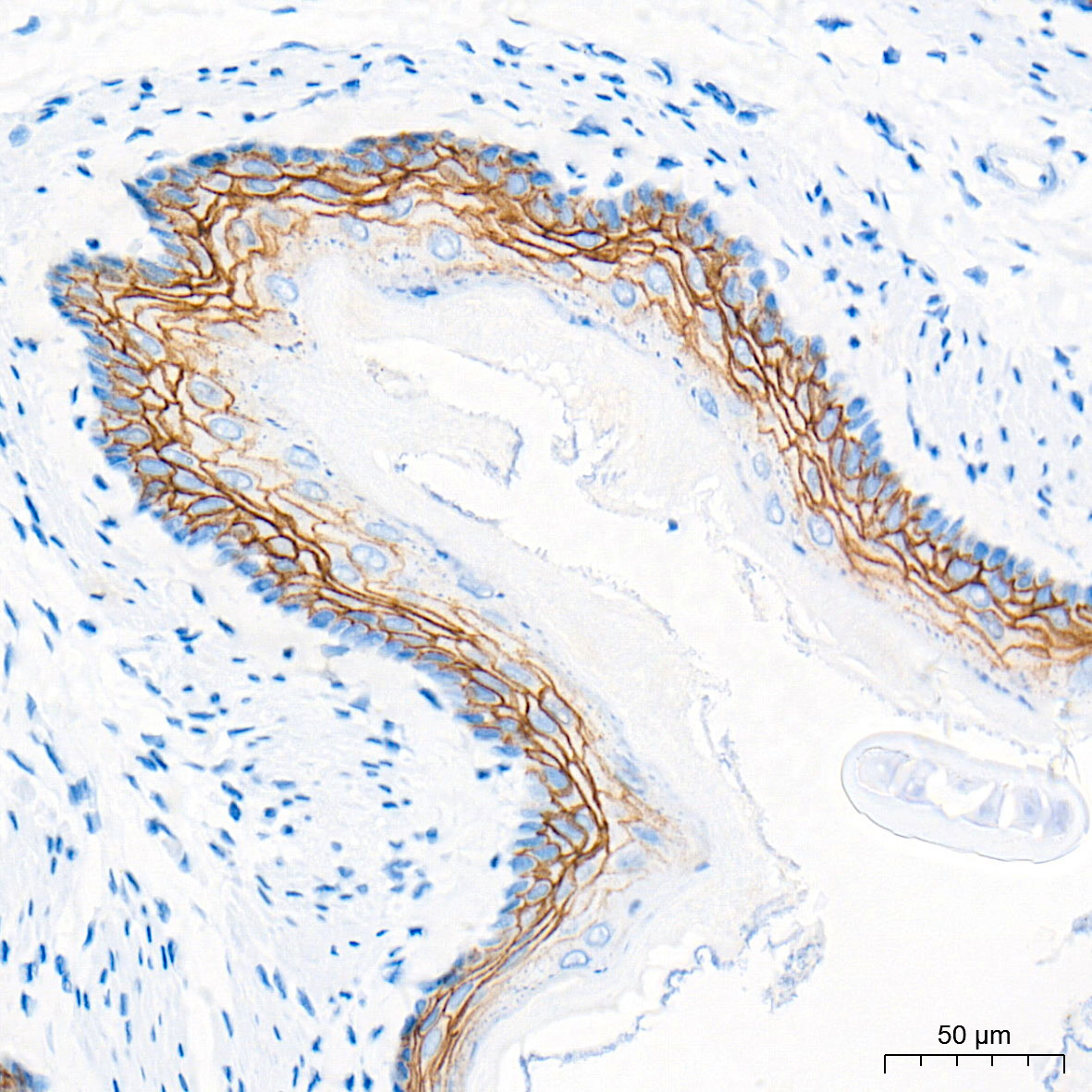
Immunohistochemistry analysis of paraffin embedded Rat esophagus tissue using Nectin-4 Rabbit mAb at a dilution of 1:500 (40x lens). High pressure antigen retrieval performed with 0.01M Tris-EDTA Buffer (pH 9.0) prior to IHC staining.ICC/IF
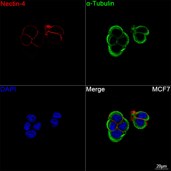
Confocal imaging of MCF7 cells using Nectin-4 Rabbit mAb (dilution 1:200) followed by a further incubation with Cy3-conjugated Goat anti-Rabbit IgG (H+L) (dilution 1:500) (Red). The cells were counterstained with α-Tubulin Mouse mAb (dilution 1:400) followed by incubation with 488-conjugated Goat Anti-Mouse IgG (H+L) Ab (dilution 1:500) (Green). DAPI was used for nuclear staining (Blue). Objective: 100x.IF-P
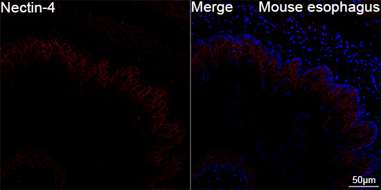
Confocal imaging of paraffin-embedded Mouse esophagus tissue using Nectin-4 Rabbit mAb (dilution 1:200) followed by a further incubation with Cy3 conjugated Goat anti-Rabbit IgG (H+L) (dilution 1:500) (Red). DAPI was used for nuclear staining (Blue). High pressure antigen retrieval performed with 0.01M Citrate Buffer (pH 6.0) prior to IF staining. Objective: 40x.| Product Name | Nectin-4 Rabbit mAb |
|---|---|
| Antibody Type | Primary Antibodies |
| Immunogen | Recombinant protein (or fragment).This information is considered to be commercially sensitive. |
| Clonality | Monoclonal |
|---|---|
| Isotype | IgG |
| Host Species | Rabbit |
| Tested Applications | ICC/IFIF-PIHCWB |
| WB:1:3000 - 1:10000 IHC:1:100 - 1:500 ICC/IF:1:100 - 1:400 IF-P:1:100 - 1:400 |
|
| Species Reactivity | HumanMouseRat |
| Concentration | 1mg/ml |
| Purification | Affinity purified |
| Gene Symbol | NECTIN4 |
|---|---|
| Gene Synonyms | LNIR PRR4 EDSS1 PVRL4 nectin-4 |
| Gene Full Name | nectin cell adhesion molecule 4 |
| Gene Summary | This gene encodes a member of the nectin family. The encoded protein contains two immunoglobulin-like (Ig-like) C2-type domains and one Ig-like V-type domain. It is involved in cell adhesion through trans-homophilic and -heterophilic interactions. It is a single-pass type I membrane protein. The soluble form is produced by proteolytic cleavage at the cell surface by the metalloproteinase ADAM17/TACE. The secreted form is found in both breast tumor cell lines and breast tumor patients. Mutations in this gene are the cause of ectodermal dysplasia-syndactyly syndrome type 1, an autosomal recessive disorder. Alternatively spliced transcript variants have been found but the full-length nature of the variant has not been determined.[provided by RefSeq, Jan 2011] |
| Alternative Names | LNIR PRR4 EDSS1 PVRL4 nectin-4 |
| Molecular Weight(MW) | 55 kDa/24 kDa(Observed band size:55-80 kDa) |
| Cellular Localization | Cell membrane, Cell junction, adherens junction, Secreted. |
WB
Western blot analysis of various lysates using Nectin-4 Rabbit mAb at 1:5000 dilution incubated overnight at 4℃. T-47D cells were treated with PNGase F (2 U/μL) at 37℃ for 1 hour. Secondary antibody: HRP-conjugated Goat anti-Rabbit IgG (H+L) at 1:10000 dilution. Lysates/proteins: 30 μg per lane. Blocking buffer: 3% nonfat dry milk in TBST. Detection: ECL Basic Kit. Negative control (NC): U-937. Exposure time: 90 s.
IHC
Immunohistochemistry analysis of paraffin embedded Human esophagus tissue using Nectin-4 Rabbit mAb at a dilution of 1:500 (40x lens). High pressure antigen retrieval performed with 0.01M Tris-EDTA Buffer (pH 9.0) prior to IHC staining.
IHC
Immunohistochemistry analysis of paraffin embedded Mouse esophagus tissue using Nectin-4 Rabbit mAb at a dilution of 1:500 (40x lens). High pressure antigen retrieval performed with 0.01M Tris-EDTA Buffer (pH 9.0) prior to IHC staining.
IHC
Immunohistochemistry analysis of paraffin embedded Rat esophagus tissue using Nectin-4 Rabbit mAb at a dilution of 1:500 (40x lens). High pressure antigen retrieval performed with 0.01M Tris-EDTA Buffer (pH 9.0) prior to IHC staining.
ICC/IF
Confocal imaging of MCF7 cells using Nectin-4 Rabbit mAb (dilution 1:200) followed by a further incubation with Cy3-conjugated Goat anti-Rabbit IgG (H+L) (dilution 1:500) (Red). The cells were counterstained with α-Tubulin Mouse mAb (dilution 1:400) followed by incubation with 488-conjugated Goat Anti-Mouse IgG (H+L) Ab (dilution 1:500) (Green). DAPI was used for nuclear staining (Blue). Objective: 100x.
IF-P
Confocal imaging of paraffin-embedded Mouse esophagus tissue using Nectin-4 Rabbit mAb (dilution 1:200) followed by a further incubation with Cy3 conjugated Goat anti-Rabbit IgG (H+L) (dilution 1:500) (Red). DAPI was used for nuclear staining (Blue). High pressure antigen retrieval performed with 0.01M Citrate Buffer (pH 6.0) prior to IF staining. Objective: 40x.| Application Notes | WB:1:3000 - 1:10000 IHC:1:100 - 1:500 ICC/IF:1:100 - 1:400 IF-P:1:100 - 1:400 |
|---|
| Form | Liquid |
|---|---|
| Storage Instructions | Store at -20℃. Avoid freeze / thaw cycles. |
| Storage Buffer | PBS with 0.09% Sodium azide, 0.05% BSA, 50% glycerol, pH7.3. |


