
WB
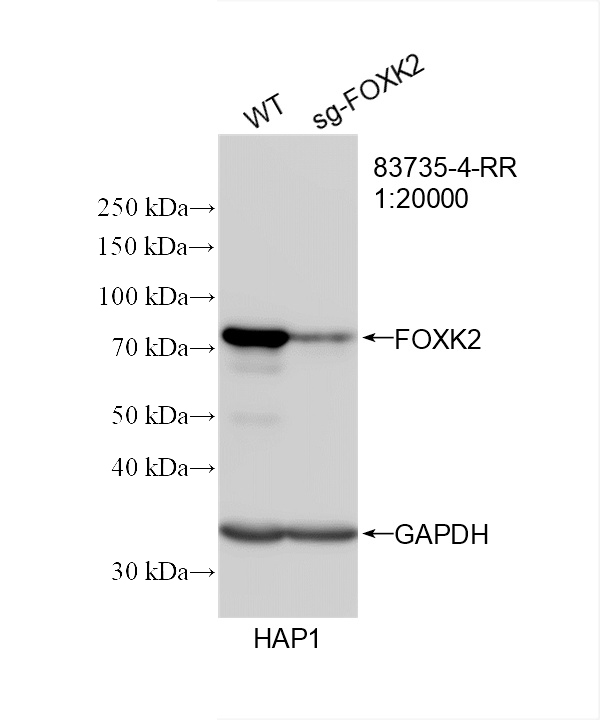
WB analysis of HAP1 using FOXK2 Recombinant monoclonal antibody. WB result of FOXK2 antibody (1:20000; room temperature for 1.5 hours) with wild-type and sg-FOXK2 transfected HAP1 cells.WB
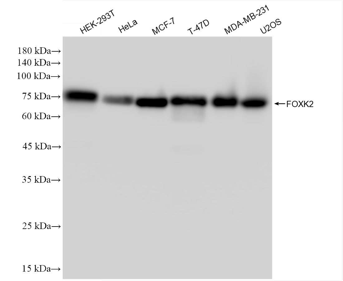
WB analysis using FOXK2 Recombinant monoclonal antibody. Various lysates were subjected to SDS PAGE followed by western blot with FOXK2 antibody at dilution of 1:20000 incubated at room temperature for 1.5 hours.IHC
IHC staining of human placenta using FOXK2 Recombinant monoclonal antibody. Immunohistochemical analysis of paraffin-embedded human placenta tissue slide using FOXK2 antibody at dilution of 1:1000 (under 10x lens). Heat mediated antigen retrieval with Tris-EDTA buffer (pH 9.0).ICC/IF
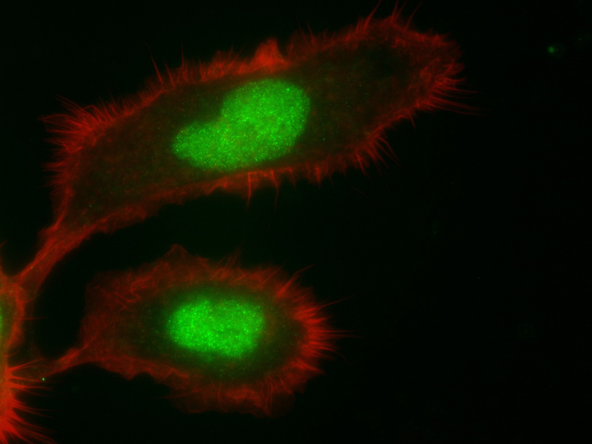
IF Staining of A431 using FOXK2 Recombinant monoclonal antibody. Immunofluorescent analysis of (4% PFA) fixed A431 cells using FOXK2 antibody at dilution of 1:250 and 488-Conjugated AffiniPure Goat Anti-Rabbit IgG(H+L), CL594-Phalloidin (red).FC
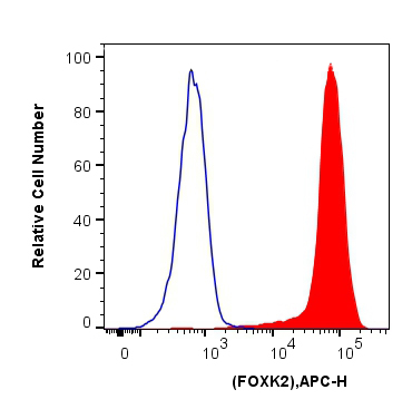
FC experiment of HEK-293T using FOXK2 Recombinant monoclonal antibody. 1x10^6 HEK-293T cells were intracellularly stained with 0.25 ug FOXK2 Recombinant antibody and APC-Conjugated AffiniPure Goat Anti-Rabbit IgG(H+L)(red), or 0.25 ug Isotype Control (blue). Cells were fixed and permeabilized with Transcription Factor Staining Buffer Kit.ChIP
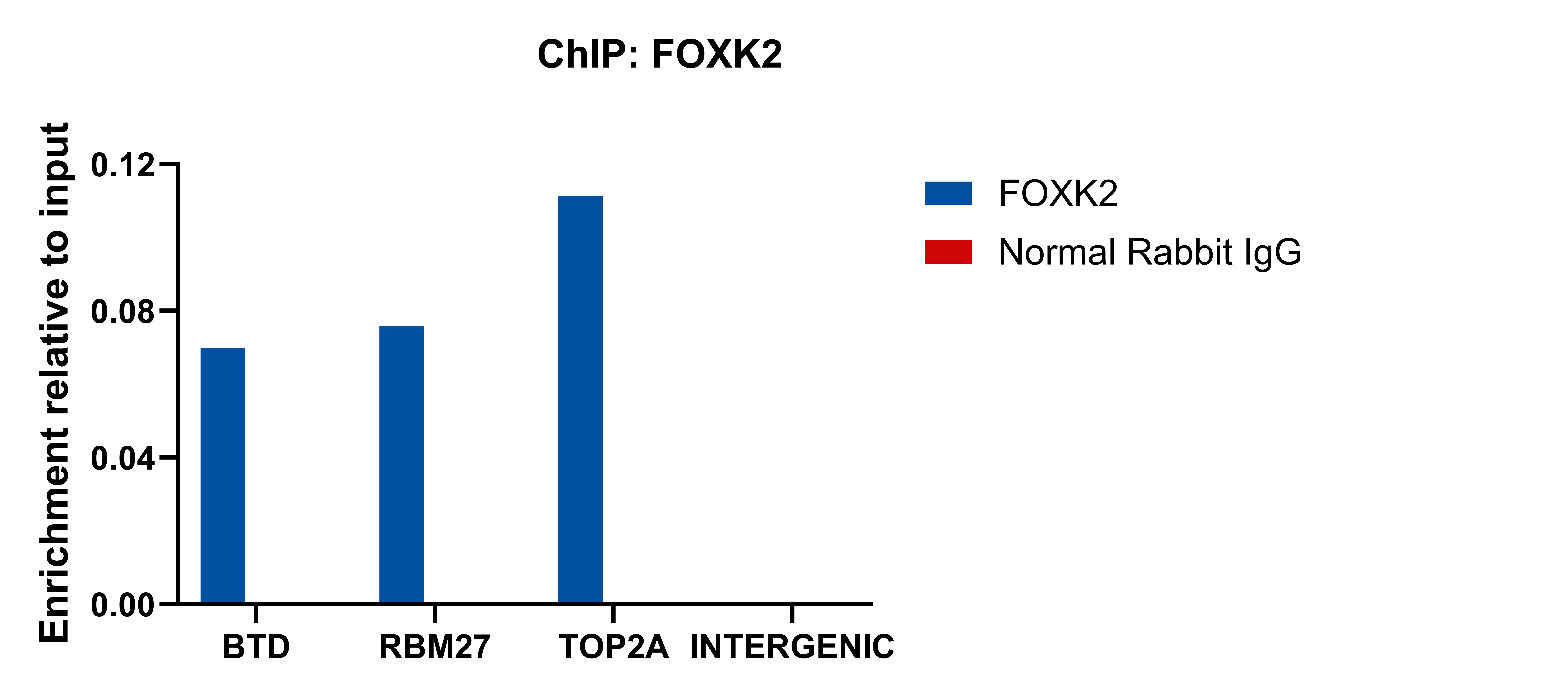
ChIP experiment of HepG2 using FOXK2 Recombinant monoclonal antibody. Chromatin was prepared from HepG2 cells. Cells were fixed with formaldehyde for 10 minutes. The ChIP was performed with 20 µg of cross-linked chromatin, 5 µg of FOXK2 or 5 ug of Normal Rabbit IgG , and 20 µl of Protein A Magarose Beads. The immunoprecipitated DNA was quantified by real-time PCR.| Product Name | FOXK2 Recombinant monoclonal antibody |
|---|---|
| Antibody Type | Primary Antibodies |
| Immunogen | Recombinant human FOXK2 protein. |
| Clonality | Monoclonal |
|---|---|
| Isotype | IgG |
| Host Species | Rabbit |
| Tested Applications | ChIPFCICC/IFIHCWB |
| WB:1:5000-1:50000 IHC:1:500-1:2000 ICC/IF:1:200-1:800 FC:0.25 ug per 10^6 cells in a 100 µl suspension ChIP:1:10-1:100 |
|
| Species Reactivity | Human |
| Concentration | 1mg/ml |
| Purification | Protein A |
| Gene Symbol | FOXK2 |
|---|---|
| Gene Synonyms | ILF ILF1 ILF-1 nGTBP |
| Gene Full Name | forkhead box K2 |
| Gene Summary | The protein encoded by this gene contains a fork head DNA binding domain. This protein can bind to the purine-rich motifs of the HIV long terminal repeat (LTR), and to the similar purine-rich motif in the interleukin 2 (IL2) promoter. It may be involved in the regulation of viral and cellular promoter elements. [provided by RefSeq, Jul 2008] |
| Alternative Names | 240692A5 forkhead box K2 Forkhead box protein K2 ILF ILF 1 |
| Molecular Weight(MW) | 69 kDa(Observed band size:69-79 kDa) |
| Cellular Localization | Cytoplasm,Nucleus. |
WB
WB analysis of HAP1 using FOXK2 Recombinant monoclonal antibody. WB result of FOXK2 antibody (1:20000; room temperature for 1.5 hours) with wild-type and sg-FOXK2 transfected HAP1 cells.
WB
WB analysis using FOXK2 Recombinant monoclonal antibody. Various lysates were subjected to SDS PAGE followed by western blot with FOXK2 antibody at dilution of 1:20000 incubated at room temperature for 1.5 hours.
IHC
IHC staining of human placenta using FOXK2 Recombinant monoclonal antibody. Immunohistochemical analysis of paraffin-embedded human placenta tissue slide using FOXK2 antibody at dilution of 1:1000 (under 10x lens). Heat mediated antigen retrieval with Tris-EDTA buffer (pH 9.0).
ICC/IF
IF Staining of A431 using FOXK2 Recombinant monoclonal antibody. Immunofluorescent analysis of (4% PFA) fixed A431 cells using FOXK2 antibody at dilution of 1:250 and 488-Conjugated AffiniPure Goat Anti-Rabbit IgG(H+L), CL594-Phalloidin (red).
FC
FC experiment of HEK-293T using FOXK2 Recombinant monoclonal antibody. 1x10^6 HEK-293T cells were intracellularly stained with 0.25 ug FOXK2 Recombinant antibody and APC-Conjugated AffiniPure Goat Anti-Rabbit IgG(H+L)(red), or 0.25 ug Isotype Control (blue). Cells were fixed and permeabilized with Transcription Factor Staining Buffer Kit.
ChIP
ChIP experiment of HepG2 using FOXK2 Recombinant monoclonal antibody. Chromatin was prepared from HepG2 cells. Cells were fixed with formaldehyde for 10 minutes. The ChIP was performed with 20 µg of cross-linked chromatin, 5 µg of FOXK2 or 5 ug of Normal Rabbit IgG , and 20 µl of Protein A Magarose Beads. The immunoprecipitated DNA was quantified by real-time PCR.| Application Notes | WB:1:5000-1:50000 IHC:1:500-1:2000 ICC/IF:1:200-1:800 FC:0.25 ug per 10^6 cells in a 100 µl suspension ChIP:1:10-1:100 |
|---|
| Form | Liquid |
|---|---|
| Storage Instructions | Store at -20°C. Stable for one year after shipment. Aliquoting is unnecessary for -20oC storage. |
| Storage Buffer | PBS with 0.02% sodium azide and 50% glycerol, pH 7.3. |


