WB
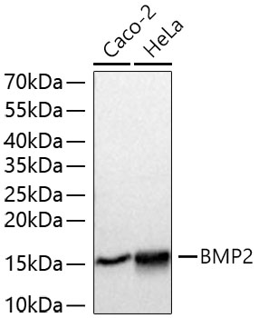
Western blot analysis of various lysates using BMP2 Rabbit pAb at 1:1000 dilution incubated at room temperature for 1.5 hours. Secondary antibody: HRP-conjugated Goat anti-Rabbit IgG (H+L) at 1:10000 dilution. Lysates/proteins: 25 μg per lane. Blocking buffer: 3% nonfat dry milk in TBST. Detection: ECL Basic Kit. Exposure time: 1 s.IHC
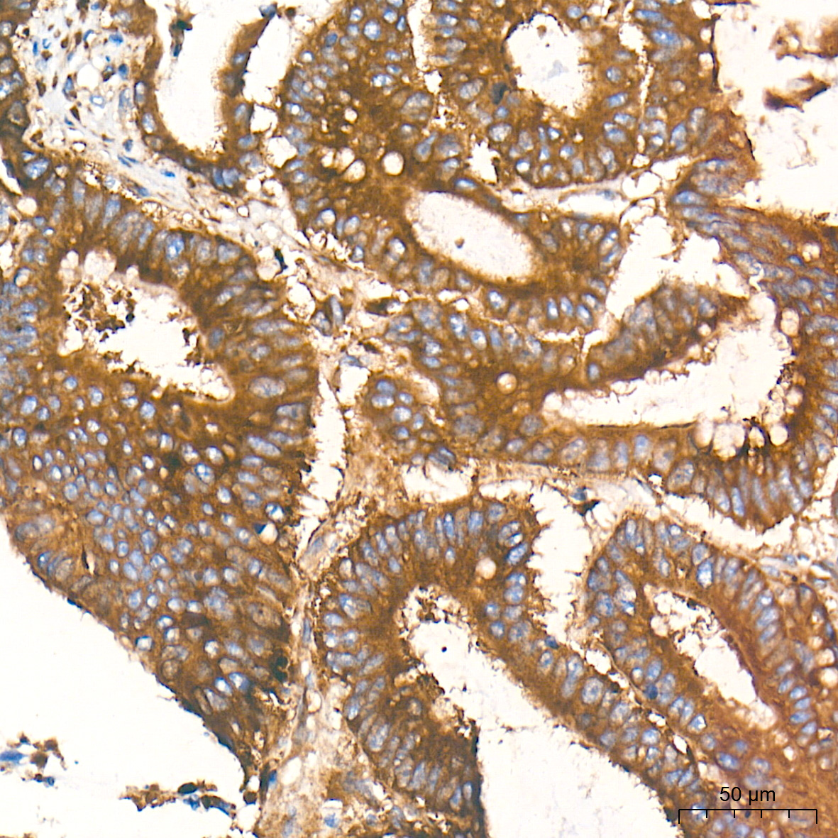
Immunohistochemistry analysis of paraffin embedded Human colon carcinoma tissue using BMP2 Rabbit pAb at a dilution of 1:200 (40x lens). High pressure antigen retrieval performed with 0.01M Tris EDTA Buffer (pH 9.0) prior to IHC staining.IHC
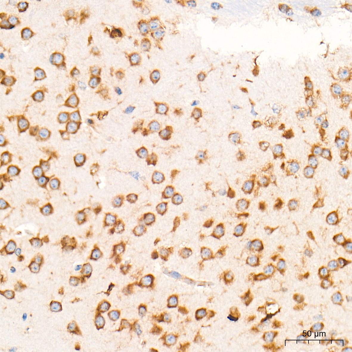
Immunohistochemistry analysis of paraffin embedded Mouse brain tissue using BMP2 Rabbit pAb at a dilution of 1:200 (40x lens). High pressure antigen retrieval performed with 0.01M Tris-EDTA Buffer (pH 9.0) prior to IHC staining.IHC
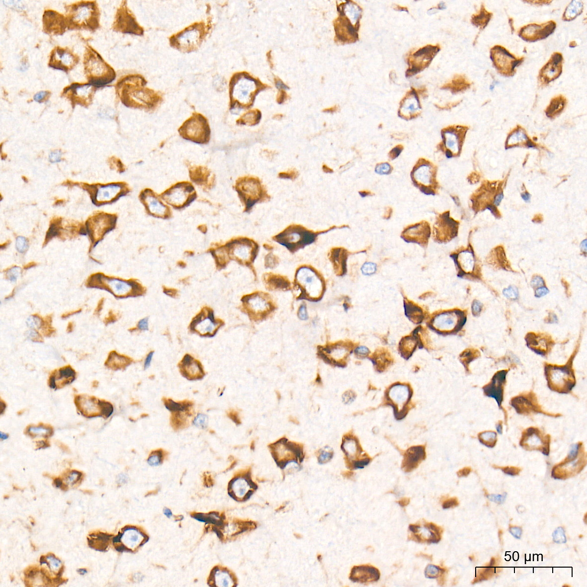
Immunohistochemistry analysis of paraffin embedded Rat brain tissue using BMP2 Rabbit pAb at a dilution of 1:200 (40x lens). High pressure antigen retrieval performed with 0.01M Tris-EDTA Buffer (pH 9.0) prior to IHC staining.ICC/IF
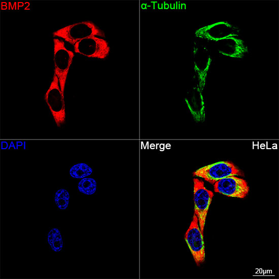
Confocal imaging of HeLa cells using BMP2 Rabbit pAb (dilution 1:200) followed by a further incubation with Cy3 conjugated Goat anti-Rabbit IgG (H+L) (dilution 1:500) (Red). The cells were counterstained with α-Tubulin Mouse mAb (dilution 1:400) followed by incubation with 488-conjugated Goat Anti-Mouse IgG (H+L) Ab (dilution 1:500) (Green). DAPI was used for nuclear staining (Blue). Objective: 100x.| Product Name | BMP2 Rabbit pAb |
|---|---|
| Antibody Type | Primary Antibodies |
| Immunogen | This information is considered to be commercially sensitive. |
| Clonality | Polyclonal |
|---|---|
| Isotype | IgG |
| Host Species | Rabbit |
| Tested Applications | ICC/IFIHCWB |
| WB:1:1000 - 1:5000 IHC:1:200 - 1:800 ICC/IF:1:200 - 1:800 |
|
| Species Reactivity | HumanMouseRat |
| Concentration | 1mg/ml |
| Purification | Affinity purified |
| Gene Symbol | BMP2 |
|---|---|
| Gene Synonyms | BDA2 BMP2A SSFSC SSFSC1 |
| Gene Full Name | bone morphogenetic protein 2 |
| Gene Summary | This gene encodes a secreted ligand of the TGF-beta (transforming growth factor-beta) superfamily of proteins. Ligands of this family bind various TGF-beta receptors leading to recruitment and activation of SMAD family transcription factors that regulate gene expression. The encoded preproprotein is proteolytically processed to generate each subunit of the disulfide-linked homodimer, which plays a role in bone and cartilage development. Duplication of a regulatory region downstream of this gene causes a form of brachydactyly characterized by a malformed index finger and second toe in human patients. [provided by RefSeq, Jul 2016] |
| Alternative Names | BDA2 BMP2A SSFSC SSFSC1 BMP2 |
| Molecular Weight(MW) | 45 kDa(Observed band size:12-45 kDa) |
| Cellular Localization | Secreted. |
WB
Western blot analysis of various lysates using BMP2 Rabbit pAb at 1:1000 dilution incubated at room temperature for 1.5 hours. Secondary antibody: HRP-conjugated Goat anti-Rabbit IgG (H+L) at 1:10000 dilution. Lysates/proteins: 25 μg per lane. Blocking buffer: 3% nonfat dry milk in TBST. Detection: ECL Basic Kit. Exposure time: 1 s.
IHC
Immunohistochemistry analysis of paraffin embedded Human colon carcinoma tissue using BMP2 Rabbit pAb at a dilution of 1:200 (40x lens). High pressure antigen retrieval performed with 0.01M Tris EDTA Buffer (pH 9.0) prior to IHC staining.
IHC
Immunohistochemistry analysis of paraffin embedded Mouse brain tissue using BMP2 Rabbit pAb at a dilution of 1:200 (40x lens). High pressure antigen retrieval performed with 0.01M Tris-EDTA Buffer (pH 9.0) prior to IHC staining.
IHC
Immunohistochemistry analysis of paraffin embedded Rat brain tissue using BMP2 Rabbit pAb at a dilution of 1:200 (40x lens). High pressure antigen retrieval performed with 0.01M Tris-EDTA Buffer (pH 9.0) prior to IHC staining.
ICC/IF
Confocal imaging of HeLa cells using BMP2 Rabbit pAb (dilution 1:200) followed by a further incubation with Cy3 conjugated Goat anti-Rabbit IgG (H+L) (dilution 1:500) (Red). The cells were counterstained with α-Tubulin Mouse mAb (dilution 1:400) followed by incubation with 488-conjugated Goat Anti-Mouse IgG (H+L) Ab (dilution 1:500) (Green). DAPI was used for nuclear staining (Blue). Objective: 100x.| Application Notes | WB:1:1000 - 1:5000 IHC:1:200 - 1:800 ICC/IF:1:200 - 1:800 |
|---|
| Form | Liquid |
|---|---|
| Storage Instructions | Store at -20℃. Avoid freeze / thaw cycles. |
| Storage Buffer | PBS, pH 7.3, containing 50% glycerol. Preserved with Proclin300 or sodium azide. May contain 0.05% BSA as specified on the Certificate of Analysis. |


