WB
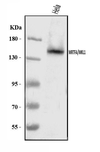
Western blot analysis of MKL1/MRTFA using anti-MKL1/MRTFA antibody. The sample well of each lane was loaded with 30 ug of sample under reducing conditions. Lane 1: Hela whole cell lysates. After electrophoresis, proteins were transferred to a membrane. Then the membrane was incubated with rabbit anti-MKL1/MRTFA antigen affinity purified polyclonal antibody at a dilution of 1:1000 and probed with a goat anti-rabbit IgG-HRP secondary antibody. The signal is developed using ECL Plus Western Blotting Substrate.ICC/IF
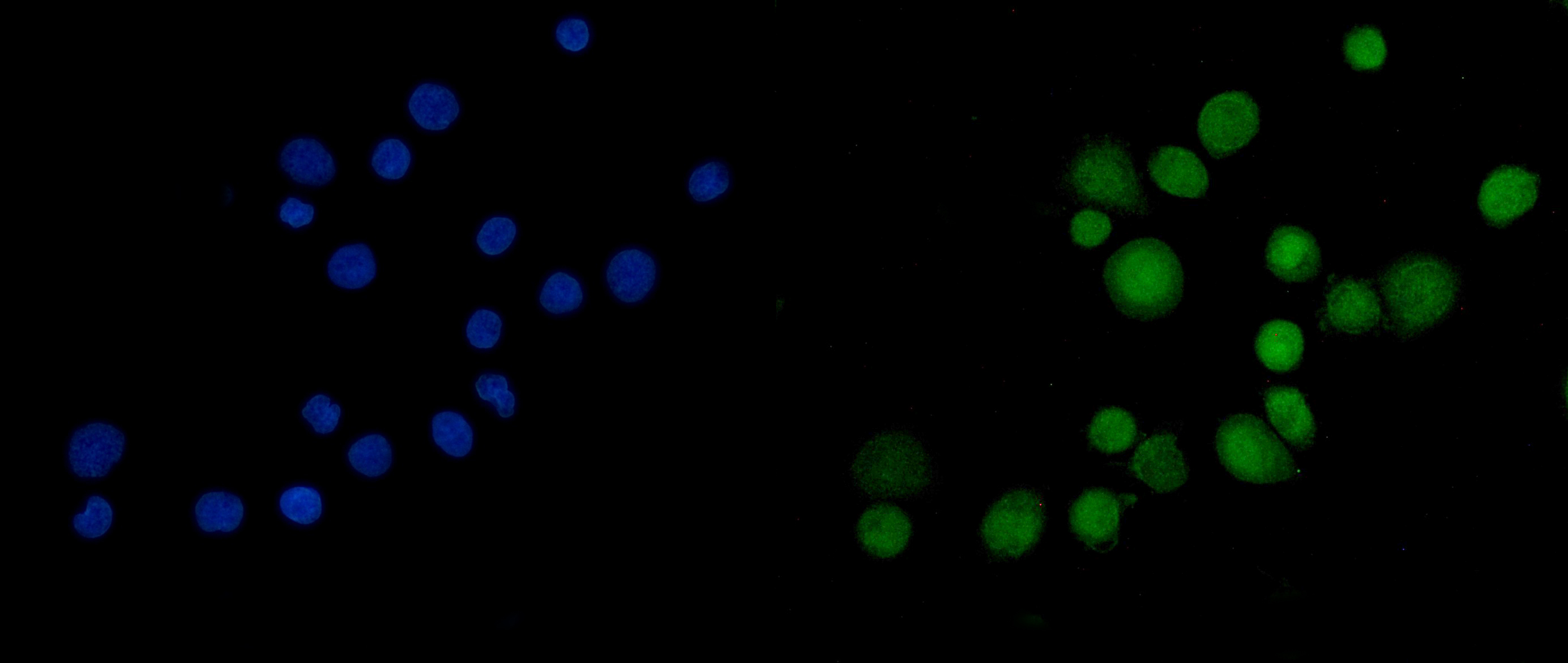
ICC/IF analysis of MKL1/MRTFA using anti-MKL1/MRTFA antibody. MKL1/MRTFA was detected in an immunocytochemical section of SiHa cells. The section was incubated with rabbit anti-MKL1/MRTFA Antibody at a dilution of 1:100. Fluoro488 Conjugated Goat Anti-Rabbit IgG (Green) was used as secondary antibody. The section was counterstained with DAPI (Blue).| Product Name | Rabbit polyclonal antibody to MKL1/MRTFA |
|---|---|
| Antibody Type | Primary Antibodies |
| Immunogen | E.coli-derived human Mkl1/MRTFA recombinant protein (Position: L192-L931). |
| Clonality | Polyclonal |
|---|---|
| Isotype | IgG |
| Host Species | Rabbit |
| Tested Applications | ICC/IFWB |
| WB:1:500-1:2000 ICC/IF:1:50-1:400 |
|
| Species Reactivity | Human |
| Concentration | 0.5mg/ml |
| Purification | Affinity purified |
| Gene Symbol | MRTFA |
|---|---|
| Gene Synonyms | MAL MKL BSAC MKL1 MRTF-A |
| Gene Full Name | myocardin related transcription factor A |
| Gene Summary | The protein encoded by this gene interacts with the transcription factor myocardin, a key regulator of smooth muscle cell differentiation. The encoded protein is predominantly nuclear and may help transduce signals from the cytoskeleton to the nucleus. This gene is involved in a specific translocation event that creates a fusion of this gene and the RNA-binding motif protein-15 gene. This translocation has been associated with acute megakaryocytic leukemia. Alternative splicing results in multiple transcript variants. [provided by RefSeq, Sep 2013] |
| Alternative Names | BSAC KIAA1438 MAL MKL/myocardin like protein 1 MKL1 MRTF A MRTFA MRTF-A |
| Molecular Weight(MW) | 99 kDa(Observed band size:145 kDa) |
| Cellular Localization | Cytoplasm,Nucleus. |
WB
Western blot analysis of MKL1/MRTFA using anti-MKL1/MRTFA antibody. The sample well of each lane was loaded with 30 ug of sample under reducing conditions. Lane 1: Hela whole cell lysates. After electrophoresis, proteins were transferred to a membrane. Then the membrane was incubated with rabbit anti-MKL1/MRTFA antigen affinity purified polyclonal antibody at a dilution of 1:1000 and probed with a goat anti-rabbit IgG-HRP secondary antibody. The signal is developed using ECL Plus Western Blotting Substrate.
ICC/IF
ICC/IF analysis of MKL1/MRTFA using anti-MKL1/MRTFA antibody. MKL1/MRTFA was detected in an immunocytochemical section of SiHa cells. The section was incubated with rabbit anti-MKL1/MRTFA Antibody at a dilution of 1:100. Fluoro488 Conjugated Goat Anti-Rabbit IgG (Green) was used as secondary antibody. The section was counterstained with DAPI (Blue).| Application Notes | WB:1:500-1:2000 ICC/IF:1:50-1:400 |
|---|
| Form | Liquid |
|---|---|
| Storage Instructions | 12 months from date of receipt,-20℃ as supplied. |
| Storage Buffer | 500 ug/ml antibody with PBS, 0.02% NaN3, 1 mg/ml BSA and 50% glycerol. |


