WB
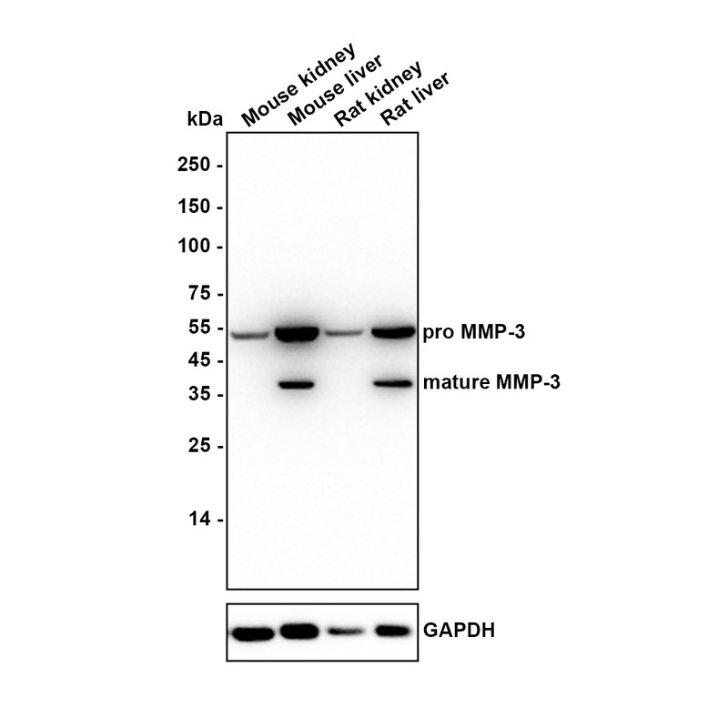
Western blot analysis of MMP-3 on different lysates with Rabbit anti-MMP-3 antibody at 1/1,000 dilution. Lysates/proteins at 40 µg/Lane. Exposure time: 1 minute; Blocking: 5% NFDM/TBST, 1 hour at room temperature. Primary antibody: 1/1,000 in 5% NFDM/TBST, overnight at 4 ℃. Secondary antibody: Goat anti-Rabbit IgG-HRP, 1/50,000 in 5% NFDM/TBST, 1 hour at room temperature.IHC
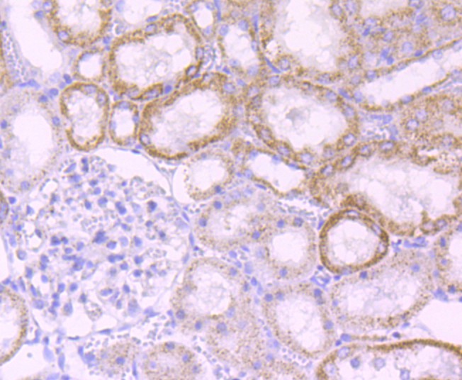
Immunohistochemical analysis of paraffin-embedded rat kidney tissue using anti-MMP-3 antibody. The section was pre-treated using heat mediated antigen retrieval with Tris-EDTA buffer (pH 8.0-8.4) for 20 minutes.The tissues were blocked in 5% BSA for 30 minutes at room temperature, washed with ddH2O and PBS, and then probed with the primary antibody (1/50) for 30 minutes at room temperature. The detection was performed using an HRP conjugated compact polymer system. DAB was used as the chromogen. Tissues were counterstained with hematoxylin and mounted with DPX.IHC
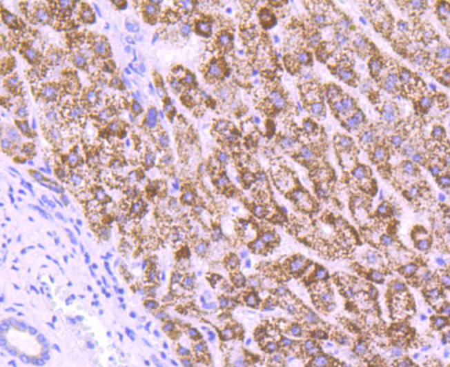
Immunohistochemical analysis of paraffin-embedded human liver tissue using anti-MMP-3 antibody. The section was pre-treated using heat mediated antigen retrieval with Tris-EDTA buffer (pH 8.0-8.4) for 20 minutes.The tissues were blocked in 5% BSA for 30 minutes at room temperature, washed with ddH2O and PBS, and then probed with the primary antibody (1/50) for 30 minutes at room temperature. The detection was performed using an HRP conjugated compact polymer system. DAB was used as the chromogen. Tissues were counterstained with hematoxylin and mounted with DPX.IHC
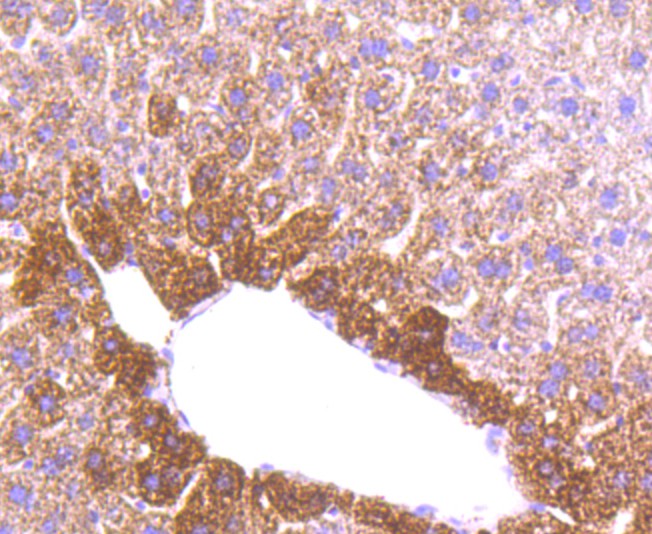
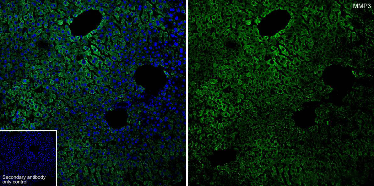
Immunohistochemical analysis of paraffin-embedded mouse liver tissue using anti-MMP-3 antibody. The section was pre-treated using heat mediated antigen retrieval with Tris-EDTA buffer (pH 8.0-8.4) for 20 minutes.The tissues were blocked in 5% BSA for 30 minutes at room temperature, washed with ddH2O and PBS, and then probed with the primary antibody (1/50) for 30 minutes at room temperature. The detection was performed using an HRP conjugated compact polymer system. DAB was used as the chromogen. Tissues were counterstained with hematoxylin and mounted with DPX.IF-P
Application: IF-Tissue. Species: Mouse. Site: liver. Sample: Paraffin-embedded section. Antibody concentration: 1/50.| Product Name | MMP-3 Recombinant Rabbit Monoclonal Antibody |
|---|---|
| Antibody Type | Primary Antibodies |
| Immunogen | Synthetic peptide within C-terminal human MMP3. |
| Clonality | Monoclonal |
|---|---|
| Isotype | IgG |
| Host Species | Rabbit |
| Tested Applications | IF-PIHCWB |
| WB:1:500-1:2000 IHC:1:50-1:200 IF-P:1:50 |
|
| Species Reactivity | HumanMouseRat |
| Concentration | 1mg/ml |
| Purification | Protein A |
| Gene Symbol | MMP3 |
|---|---|
| Gene Synonyms | SL-1 STMY STR1 CHDS6 MMP-3 STMY1 |
| Gene Full Name | matrix metallopeptidase 3 |
| Gene Summary | Proteins of the matrix metalloproteinase (MMP) family are involved in the breakdown of extracellular matrix in normal physiological processes, such as embryonic development, reproduction, and tissue remodeling, as well as in disease processes, such as arthritis and metastasis. Most MMP's are secreted as inactive proproteins which are activated when cleaved by extracellular proteinases. This gene encodes an enzyme which degrades fibronectin, laminin, collagens III, IV, IX, and X, and cartilage proteoglycans. The enzyme is thought to be involved in wound repair, progression of atherosclerosis, and tumor initiation. The gene is part of a cluster of MMP genes which localize to chromosome 11q22.3. [provided by RefSeq, Jul 2008] |
| Alternative Names | CHDS6 antibody Matrix metalloproteinase 3 antibody Matrix metalloproteinase-3 antibody MGC126102 antibody MGC126103 antibody MGC126104 antibody MMP 3 antibody MMP-3 antibody MMP3 antibody MMP3_HUMAN antibody Proteoglycanase antibody SL-1 antibody SL1 antibody STMY antibody STMY1 antibody STR1 antibody Stromelysin 1 antibody Stromelysin-1 antibody Transin 1 antibody Transin-1 antibody |
| Molecular Weight(MW) | 54 kDa(Observed band size:54/41 kDa) |
| Cellular Localization | extracellular matrix. |
WB
Western blot analysis of MMP-3 on different lysates with Rabbit anti-MMP-3 antibody at 1/1,000 dilution. Lysates/proteins at 40 µg/Lane. Exposure time: 1 minute; Blocking: 5% NFDM/TBST, 1 hour at room temperature. Primary antibody: 1/1,000 in 5% NFDM/TBST, overnight at 4 ℃. Secondary antibody: Goat anti-Rabbit IgG-HRP, 1/50,000 in 5% NFDM/TBST, 1 hour at room temperature.
IHC
Immunohistochemical analysis of paraffin-embedded rat kidney tissue using anti-MMP-3 antibody. The section was pre-treated using heat mediated antigen retrieval with Tris-EDTA buffer (pH 8.0-8.4) for 20 minutes.The tissues were blocked in 5% BSA for 30 minutes at room temperature, washed with ddH2O and PBS, and then probed with the primary antibody (1/50) for 30 minutes at room temperature. The detection was performed using an HRP conjugated compact polymer system. DAB was used as the chromogen. Tissues were counterstained with hematoxylin and mounted with DPX.
IHC
Immunohistochemical analysis of paraffin-embedded human liver tissue using anti-MMP-3 antibody. The section was pre-treated using heat mediated antigen retrieval with Tris-EDTA buffer (pH 8.0-8.4) for 20 minutes.The tissues were blocked in 5% BSA for 30 minutes at room temperature, washed with ddH2O and PBS, and then probed with the primary antibody (1/50) for 30 minutes at room temperature. The detection was performed using an HRP conjugated compact polymer system. DAB was used as the chromogen. Tissues were counterstained with hematoxylin and mounted with DPX.
IHC
Immunohistochemical analysis of paraffin-embedded mouse liver tissue using anti-MMP-3 antibody. The section was pre-treated using heat mediated antigen retrieval with Tris-EDTA buffer (pH 8.0-8.4) for 20 minutes.The tissues were blocked in 5% BSA for 30 minutes at room temperature, washed with ddH2O and PBS, and then probed with the primary antibody (1/50) for 30 minutes at room temperature. The detection was performed using an HRP conjugated compact polymer system. DAB was used as the chromogen. Tissues were counterstained with hematoxylin and mounted with DPX.
IF-P
Application: IF-Tissue. Species: Mouse. Site: liver. Sample: Paraffin-embedded section. Antibody concentration: 1/50.| Application Notes | WB:1:500-1:2000 IHC:1:50-1:200 IF-P:1:50 |
|---|
| Form | Liquid |
|---|---|
| Storage Instructions | Shipped at 4℃. Store at +4℃ short term (1-2 weeks). Store at -20℃ long term. |
| Storage Buffer | 1*TBS (pH7.4), 0.05% BSA, 40% Glycerol. Preservative: 0.05% Sodium Azide. |


