WB
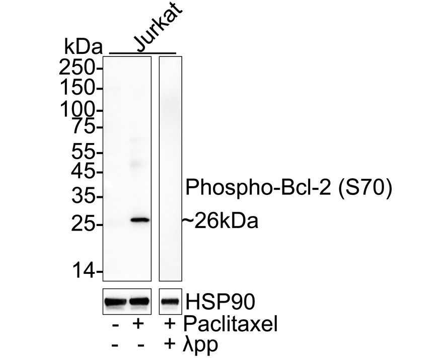
Cell treatment (CT) Western blot analysis of Phospho-Bcl-2 (S70) on different lysates with Rabbit anti-Phospho-Bcl-2 (S70) antibody at 1/2,000 dilution. Lysates/proteins at 20 µg/Lane. Exposure time: 1 minute 15 seconds; 4-20% SDS-PAGE gel. Proteins were transferred to a PVDF membrane and blocked with 5% NFDM/TBST for 1 hour at room temperature. The primary antibody at 1/2,000 dilution was used in 5% NFDM/TBST at room temperature for 2 hours. Goat Anti-Rabbit IgG - HRP Secondary Antibody at 1/50,000 dilution was used for 1 hour at room temperature.ICC/IF
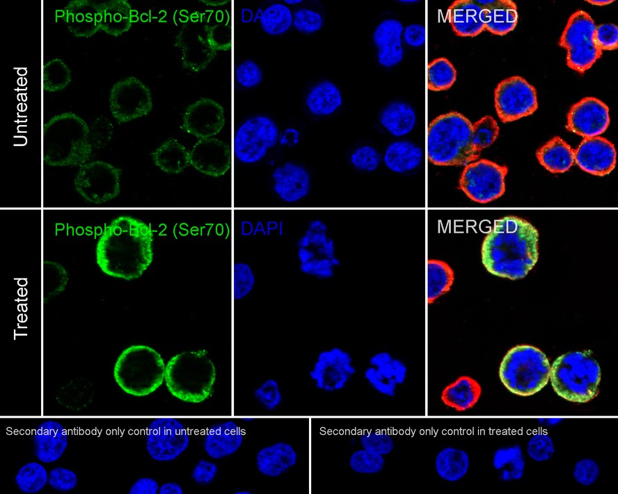
Cell treatment (CT). Immunocytochemistry analysis of Jurkat cells untreated / treated with 1μM paclitaxel overnight labeling Phospho-Bcl-2 (S70) with Rabbit anti-Phospho-Bcl-2 (S70) antibody at 1/500 dilution. Cells were fixed in 4% paraformaldehyde for 15 minutes at room temperature, permeabilized with 0.1% Triton X-100 in PBS for 15 minutes at room temperature, then blocked with 1% BSA in 10% negative goat serum for 1 hour at room temperature. Cells were then incubated with Rabbit anti-Phospho-Bcl-2 (S70) antibody at 1/500 dilution in 1% BSA in PBST overnight at 4 ℃. Goat Anti-Rabbit IgG H&L (488) was used as the secondary antibody at 1/1,000 dilution. PBS instead of the primary antibody was used as the secondary antibody only control. Nuclear DNA was labelled in blue with DAPI. Beta tubulin (red) was stained at 1/100 dilution overnight at +4℃. Goat Anti-Mouse IgG H&L (594) was used as the secondary antibody at 1/1,000 dilution.FC
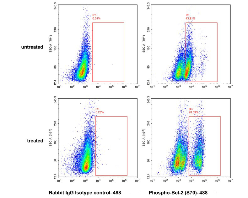
Cell treatment (CT). Flow cytometric analysis of untreated Jurkat cells (top) / Jurkat cells treated with 1μM paclitaxel overnight (bottom) labeling Phospho-Bcl-2 (S70). Cells were fixed and permeabilized. Then stained with the primary antibody (1/10,000) (right) compared with Rabbit IgG Isotype Control (1μg/mL, left). After incubation of the primary antibody at +4℃ for an hour, the cells were stained with a 488 conjugate-Goat anti-Rabbit IgG Secondary antibody at 1/1,000 dilution for 30 minutes at +4℃.| Product Name | Phospho-Bcl-2 (S70) Recombinant Rabbit Monoclonal Antibody |
|---|---|
| Antibody Type | Primary Antibodies |
| Immunogen | Synthetic phospho-peptide corresponding to residues surrounding Ser70 of Human Bcl-2. |
| Modification | p-S70 |
| Clonality | Monoclonal |
|---|---|
| Isotype | IgG |
| Host Species | Rabbit |
| Tested Applications | FCICC/IFWB |
| WB:1:2000 ICC/IF:1:500 FC:1:10000 |
|
| Species Reactivity | Human |
| Concentration | 1mg/ml |
| Purification | Protein A |
| Gene Symbol | BCL2 |
|---|---|
| Gene Synonyms | Bcl-2 PPP1R50 |
| Gene Full Name | BCL2 apoptosis regulator |
| Gene Summary | This gene encodes an integral outer mitochondrial membrane protein that blocks the apoptotic death of some cells such as lymphocytes. Constitutive expression of BCL2, such as in the case of translocation of BCL2 to Ig heavy chain locus, is thought to be the cause of follicular lymphoma. Alternative splicing results in multiple transcript variants. [provided by RefSeq, Feb 2016] |
| Alternative Names | Apoptosis regulator Bcl 2 antibody Apoptosis regulator Bcl-2 antibody Apoptosis regulator Bcl2 antibody AW986256 antibody B cell CLL/lymphoma 2 antibody B cell leukemia/lymphoma 2 antibody Bcl-2 antibody Bcl2 antibody BCL2_HUMAN antibody C430015F12Rik antibody D630044D05Rik antibody D830018M01Rik antibody Leukemia/lymphoma B-cell 2 antibody Oncogene B-cell leukemia 2 antibody PPP1R50 antibody Protein phosphatase 1 regulatory subunit 50 antibody Bcl 2 antibody |
| Molecular Weight(MW) | 26 kDa |
| Cellular Localization | Mitochondrion outer membrane, Nucleus membrane, Endoplasmic reticulum membrane, Cytoplasm. |
WB
Cell treatment (CT) Western blot analysis of Phospho-Bcl-2 (S70) on different lysates with Rabbit anti-Phospho-Bcl-2 (S70) antibody at 1/2,000 dilution. Lysates/proteins at 20 µg/Lane. Exposure time: 1 minute 15 seconds; 4-20% SDS-PAGE gel. Proteins were transferred to a PVDF membrane and blocked with 5% NFDM/TBST for 1 hour at room temperature. The primary antibody at 1/2,000 dilution was used in 5% NFDM/TBST at room temperature for 2 hours. Goat Anti-Rabbit IgG - HRP Secondary Antibody at 1/50,000 dilution was used for 1 hour at room temperature.
ICC/IF
Cell treatment (CT). Immunocytochemistry analysis of Jurkat cells untreated / treated with 1μM paclitaxel overnight labeling Phospho-Bcl-2 (S70) with Rabbit anti-Phospho-Bcl-2 (S70) antibody at 1/500 dilution. Cells were fixed in 4% paraformaldehyde for 15 minutes at room temperature, permeabilized with 0.1% Triton X-100 in PBS for 15 minutes at room temperature, then blocked with 1% BSA in 10% negative goat serum for 1 hour at room temperature. Cells were then incubated with Rabbit anti-Phospho-Bcl-2 (S70) antibody at 1/500 dilution in 1% BSA in PBST overnight at 4 ℃. Goat Anti-Rabbit IgG H&L (488) was used as the secondary antibody at 1/1,000 dilution. PBS instead of the primary antibody was used as the secondary antibody only control. Nuclear DNA was labelled in blue with DAPI. Beta tubulin (red) was stained at 1/100 dilution overnight at +4℃. Goat Anti-Mouse IgG H&L (594) was used as the secondary antibody at 1/1,000 dilution.
FC
Cell treatment (CT). Flow cytometric analysis of untreated Jurkat cells (top) / Jurkat cells treated with 1μM paclitaxel overnight (bottom) labeling Phospho-Bcl-2 (S70). Cells were fixed and permeabilized. Then stained with the primary antibody (1/10,000) (right) compared with Rabbit IgG Isotype Control (1μg/mL, left). After incubation of the primary antibody at +4℃ for an hour, the cells were stained with a 488 conjugate-Goat anti-Rabbit IgG Secondary antibody at 1/1,000 dilution for 30 minutes at +4℃.| Application Notes | WB:1:2000 ICC/IF:1:500 FC:1:10000 |
|---|
| Form | Liquid |
|---|---|
| Storage Instructions | Shipped at 4℃. Store at +4℃ short term (1-2 weeks). Store at -20℃ long term. |
| Storage Buffer | 1*PBS (pH7.4), 0.1% BSA, 40% Glycerol, 0.2% Proclean 950. |


