WB
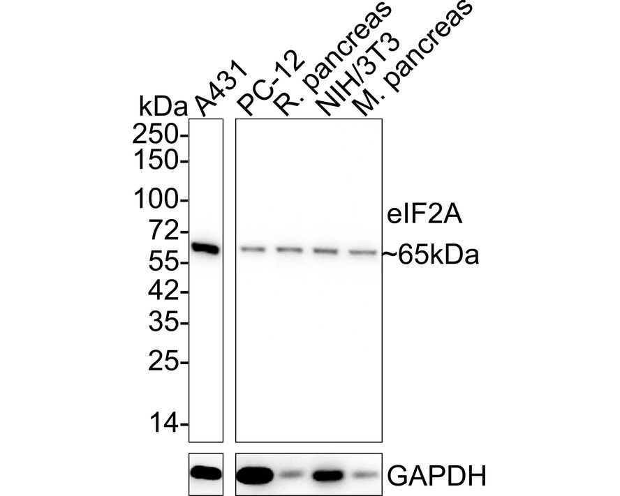
Western blot analysis of eIF2A on different lysates with Rabbit anti-eIF2A antibody at 1/1,000 dilution. Exposure time: 24 seconds; 4-20% SDS-PAGE gel. Proteins were transferred to a PVDF membrane and blocked with 5% NFDM/TBST for 1 hour at room temperature. The primary antibody at 1/1,000 dilution was used in 5% NFDM/TBST at 4℃ overnight. Goat Anti-Rabbit IgG - HRP Secondary Antibody at 1/50,000 dilution was used for 1 hour at room temperature.IHC
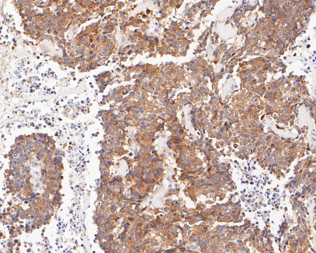
Immunohistochemical analysis of paraffin-embedded human lung carcinoma tissue using anti-eIF2A antibody. The section was pre-treated using heat mediated antigen retrieval with Tris-EDTA buffer (pH 8.0-8.4) for 20 minutes.The tissues were blocked in 5% BSA for 30 minutes at room temperature, washed with ddH2O and PBS, and then probed with the primary antibody (1/200) for 30 minutes at room temperature. The detection was performed using an HRP conjugated compact polymer system. DAB was used as the chromogen. Tissues were counterstained with hematoxylin and mounted with DPX.FC
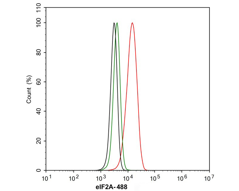
Flow cytometric analysis of NIH/3T3 cells labeling eIF2A. Cells were fixed and permeabilized. Then stained with the primary antibody (1μg/mL) (red) compared with Rabbit IgG Isotype Control (green). After incubation of the primary antibody at +4℃ for an hour, the cells were stained with a 488 conjugate-Goat anti-Rabbit IgG Secondary antibody at 1/1,000 dilution for 30 minutes at +4℃. Unlabelled sample was used as a control (cells without incubation with primary antibody; black).FC
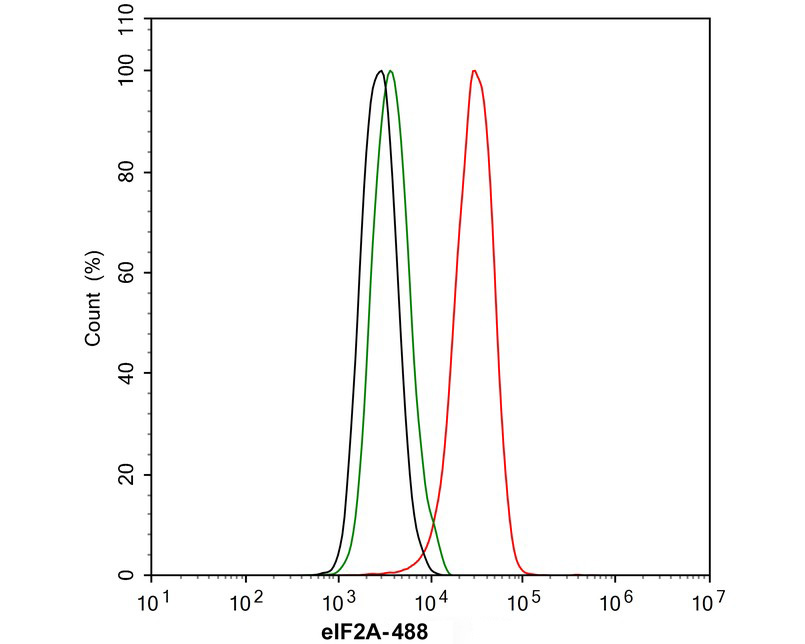
Flow cytometric analysis of PC-12 cells labeling eIF2A. Cells were fixed and permeabilized. Then stained with the primary antibody (1μg/mL) (red) compared with Rabbit IgG Isotype Control (green). After incubation of the primary antibody at +4℃ for an hour, the cells were stained with a 488 conjugate-Goat anti-Rabbit IgG Secondary antibody at 1/1,000 dilution for 30 minutes at +4℃. Unlabelled sample was used as a control (cells without incubation with primary antibody; black).| Product Name | eIF2A Recombinant Rabbit Monoclonal Antibody |
|---|---|
| Antibody Type | Primary Antibodies |
| Immunogen | Recombinant protein within human EIF2A aa 435-585. |
| Clonality | Monoclonal |
|---|---|
| Isotype | IgG |
| Host Species | Rabbit |
| Tested Applications | FCIHCWB |
| WB:1:1000 IHC:1:50-1:200 FC:1:1000 |
|
| Species Reactivity | HumanMouseRat |
| Concentration | 1mg/ml |
| Purification | Protein A |
| Gene Symbol | EIF2A |
|---|---|
| Gene Synonyms | CDA02 EIF-2A MST089 MSTP004 MSTP089 |
| Gene Full Name | eukaryotic translation initiation factor 2A |
| Gene Summary | This gene encodes a eukaryotic translation initiation factor that catalyzes the formation of puromycin-sensitive 80 S preinitiation complexes and the poly(U)-directed synthesis of polyphenylalanine at low concentrations of Mg2+. This gene should not be confused with eIF2-alpha (EIF2S1, Gene ID: 1965), the alpha subunit of the eIF2 translation initiation complex. Although both of these proteins function in binding initiator tRNA to the 40 S ribosomal subunit, the encoded protein does so in a codon-dependent manner, whereas eIF2 complex requires GTP. Alternative splicing of this gene results in multiple transcript variants encoding different isoforms. [provided by RefSeq, Jan 2016] |
| Alternative Names | 65 kDa eukaryotic translation initiation factor 2A antibody CDA 02 antibody CDA02 antibody EIF 2 antibody EIF 2A antibody eIF-2A antibody EIF2 antibody eif2a antibody EIF2A_HUMAN antibody Eukaryotic translation initiation factor 2A 65kDa antibody Eukaryotic translation initiation factor 2A antibody MST089 antibody MSTP004 antibody MSTP089 antibody |
| Molecular Weight(MW) | 65 kDa |
| Cellular Localization | Cytoplasm, extracellular region or secreted. |
WB
Western blot analysis of eIF2A on different lysates with Rabbit anti-eIF2A antibody at 1/1,000 dilution. Exposure time: 24 seconds; 4-20% SDS-PAGE gel. Proteins were transferred to a PVDF membrane and blocked with 5% NFDM/TBST for 1 hour at room temperature. The primary antibody at 1/1,000 dilution was used in 5% NFDM/TBST at 4℃ overnight. Goat Anti-Rabbit IgG - HRP Secondary Antibody at 1/50,000 dilution was used for 1 hour at room temperature.
IHC
Immunohistochemical analysis of paraffin-embedded human lung carcinoma tissue using anti-eIF2A antibody. The section was pre-treated using heat mediated antigen retrieval with Tris-EDTA buffer (pH 8.0-8.4) for 20 minutes.The tissues were blocked in 5% BSA for 30 minutes at room temperature, washed with ddH2O and PBS, and then probed with the primary antibody (1/200) for 30 minutes at room temperature. The detection was performed using an HRP conjugated compact polymer system. DAB was used as the chromogen. Tissues were counterstained with hematoxylin and mounted with DPX.
FC
Flow cytometric analysis of NIH/3T3 cells labeling eIF2A. Cells were fixed and permeabilized. Then stained with the primary antibody (1μg/mL) (red) compared with Rabbit IgG Isotype Control (green). After incubation of the primary antibody at +4℃ for an hour, the cells were stained with a 488 conjugate-Goat anti-Rabbit IgG Secondary antibody at 1/1,000 dilution for 30 minutes at +4℃. Unlabelled sample was used as a control (cells without incubation with primary antibody; black).
FC
Flow cytometric analysis of PC-12 cells labeling eIF2A. Cells were fixed and permeabilized. Then stained with the primary antibody (1μg/mL) (red) compared with Rabbit IgG Isotype Control (green). After incubation of the primary antibody at +4℃ for an hour, the cells were stained with a 488 conjugate-Goat anti-Rabbit IgG Secondary antibody at 1/1,000 dilution for 30 minutes at +4℃. Unlabelled sample was used as a control (cells without incubation with primary antibody; black).| Application Notes | WB:1:1000 IHC:1:50-1:200 FC:1:1000 |
|---|
| Form | Liquid |
|---|---|
| Storage Instructions | Shipped at 4℃. Store at +4℃ short term (1-2 weeks). Store at -20℃ long term. |
| Storage Buffer | 1*TBS (pH7.4), 0.05% BSA, 40% Glycerol. Preservative: 0.05% Sodium Azide. |


