WB
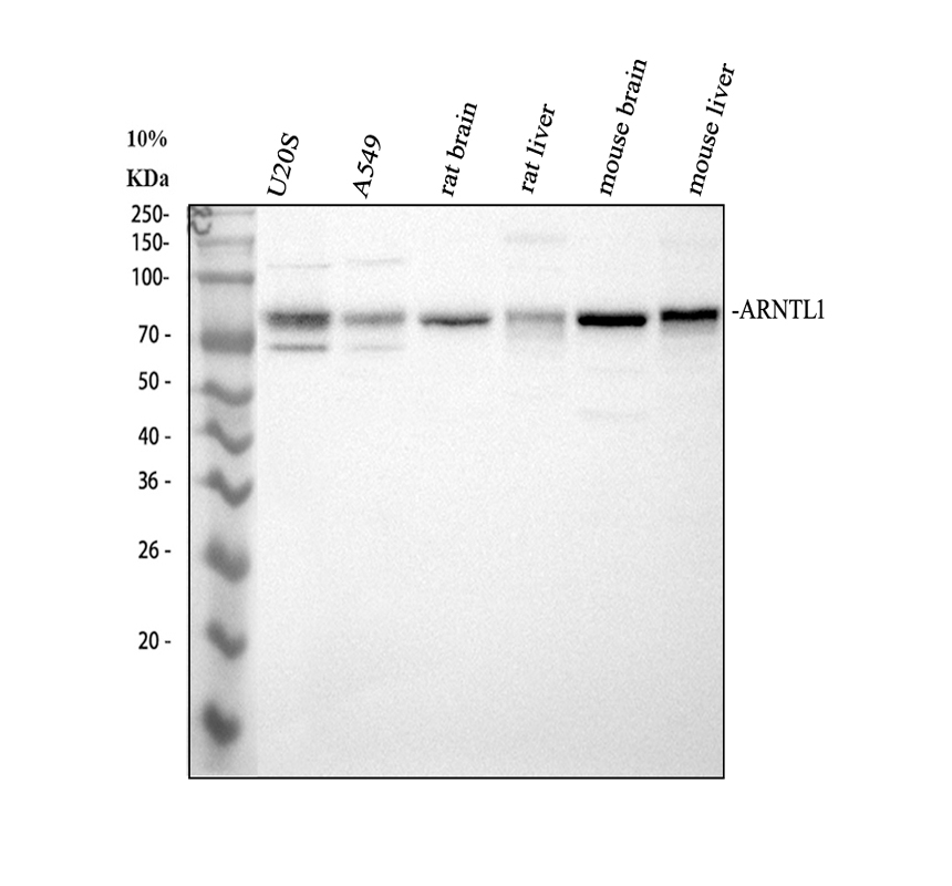
Western blot analysis of ANTXR1 using anti-ANTXR1 antibody. The sample well of each lane was loaded with 30 ug of sample under reducing conditions. After electrophoresis, proteins were transferred to a membrane. Then the membrane was incubated with rabbit anti-ANTXR1 antigen affinity purified polyclonal antibody at a dilution of 1:1000 and probed with a goat anti-rabbit IgG-HRP secondary antibody. The signal is developed using ECL Plus Western Blotting Substrate.IHC
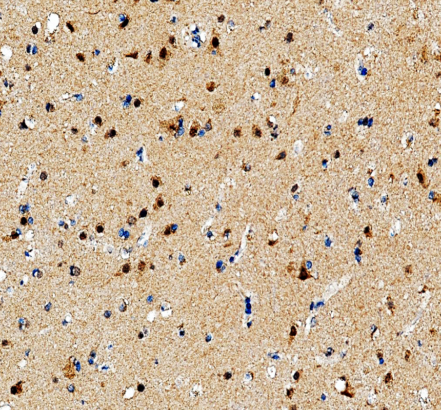
IHC analysis of ANTXR1 using anti-ANTXR1 antibody. ANTXR1 was detected in a paraffin-embedded section of human brain tissue. The tissue section was incubated with rabbit anti-ANTXR1 Antibody at a dilution of 1:200 and developed using HRP Conjugated Rabbit IgG Super Vision Assay Kit with DAB as the chromogen.IHC
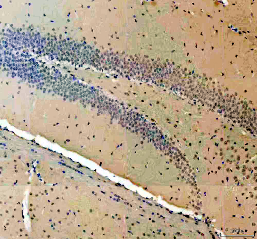
IHC analysis of ANTXR1 using anti-ANTXR1 antibody. ANTXR1 was detected in a paraffin-embedded section of mouse brain tissue. The tissue section was incubated with rabbit anti-ANTXR1 Antibody at a dilution of 1:200 and developed using HRP Conjugated Rabbit IgG Super Vision Assay Kit with DAB as the chromogen.IHC
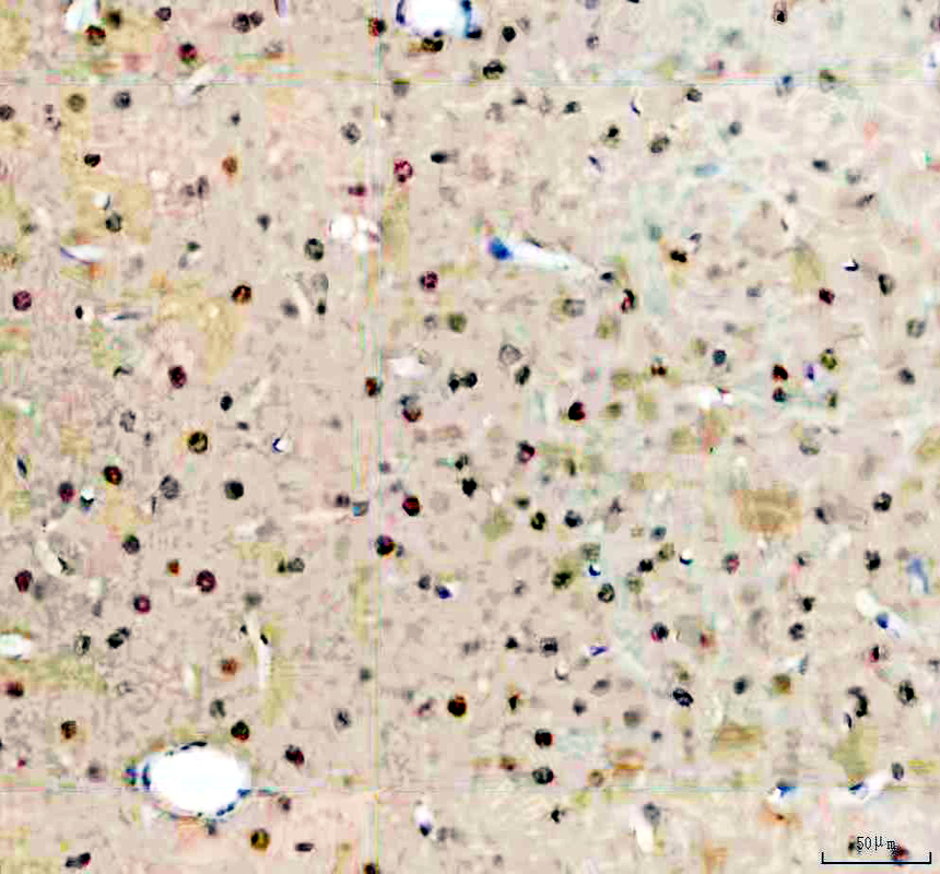
IHC analysis of ANTXR1 using anti-ANTXR1 antibody. ANTXR1 was detected in a paraffin-embedded section of rat brain tissue. The tissue section was incubated with rabbit anti-ANTXR1 Antibody at a dilution of 1:200 and developed using HRP Conjugated Rabbit IgG Super Vision Assay Kit with DAB as the chromogen.ICC/IF
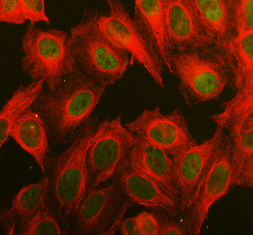
ICC/IF analysis of ANTXR1 using anti-ANTXR1 antibody and anti-Alpha Tubulin antibody. ANTXR1 was detected in an immunocytochemical section of U2OS cells. The section was incubated with rabbit anti-ANTXR1 Antibody at a dilution of 1:100. Fluoro488-conjugated Anti-rabbit IgG Secondary Antibody (green) and Cy3-conjugated Anti-mouse IgG Secondary Antibody (red) were used as secondary antibody.| Product Name | Rabbit polyclonal antibody to ARNTL |
|---|---|
| Antibody Type | Primary Antibodies |
| Immunogen | E.coli-derived human BMAL1/ARNTL recombinant protein (Position: R116-E447). |
| Clonality | Polyclonal |
|---|---|
| Isotype | IgG |
| Host Species | Rabbit |
| Tested Applications | ICC/IFIHCWB |
| WB:1:1000-5000 IHC:1:50-1:400 ICC/IF:1:50-1:400 |
|
| Species Reactivity | HumanMouseRat |
| Concentration | 0.5mg/ml |
| Purification | Affinity purified |
| Gene Symbol | BMAL1 |
|---|---|
| Gene Synonyms | TIC JAP3 MOP3 ARNTL PASD3 ARNTL1 BMAL1c bHLHe5 |
| Gene Full Name | basic helix-loop-helix ARNT like 1 |
| Gene Summary | The protein encoded by this gene is a basic helix-loop-helix protein that forms a heterodimer with CLOCK. This heterodimer binds E-box enhancer elements upstream of Period (PER1, PER2, PER3) and Cryptochrome (CRY1, CRY2) genes and activates transcription of these genes. PER and CRY proteins heterodimerize and repress their own transcription by interacting in a feedback loop with CLOCK/ARNTL complexes. Defects in this gene have been linked to infertility, problems with gluconeogenesis and lipogenesis, and altered sleep patterns. The protein regulates interferon-stimulated gene expression and is an important factor in viral infection, including COVID-19. [provided by RefSeq, Oct 2021] |
| Alternative Names | ARNT3 ARNTL bHLH PAS protein JAP3 bHLHe5 BMAL1 BMAL1c Brain and muscle ARNT like 1 JAP3 Member of PAS protein 3 MOP3 PASD3 TIC |
| Molecular Weight(MW) | 69 kDa(Observed band size:75 kDa) |
| Cellular Localization | Cytoplasm,Nucleus. |
WB
Western blot analysis of ANTXR1 using anti-ANTXR1 antibody. The sample well of each lane was loaded with 30 ug of sample under reducing conditions. After electrophoresis, proteins were transferred to a membrane. Then the membrane was incubated with rabbit anti-ANTXR1 antigen affinity purified polyclonal antibody at a dilution of 1:1000 and probed with a goat anti-rabbit IgG-HRP secondary antibody. The signal is developed using ECL Plus Western Blotting Substrate.
IHC
IHC analysis of ANTXR1 using anti-ANTXR1 antibody. ANTXR1 was detected in a paraffin-embedded section of human brain tissue. The tissue section was incubated with rabbit anti-ANTXR1 Antibody at a dilution of 1:200 and developed using HRP Conjugated Rabbit IgG Super Vision Assay Kit with DAB as the chromogen.
IHC
IHC analysis of ANTXR1 using anti-ANTXR1 antibody. ANTXR1 was detected in a paraffin-embedded section of mouse brain tissue. The tissue section was incubated with rabbit anti-ANTXR1 Antibody at a dilution of 1:200 and developed using HRP Conjugated Rabbit IgG Super Vision Assay Kit with DAB as the chromogen.
IHC
IHC analysis of ANTXR1 using anti-ANTXR1 antibody. ANTXR1 was detected in a paraffin-embedded section of rat brain tissue. The tissue section was incubated with rabbit anti-ANTXR1 Antibody at a dilution of 1:200 and developed using HRP Conjugated Rabbit IgG Super Vision Assay Kit with DAB as the chromogen.
ICC/IF
ICC/IF analysis of ANTXR1 using anti-ANTXR1 antibody and anti-Alpha Tubulin antibody. ANTXR1 was detected in an immunocytochemical section of U2OS cells. The section was incubated with rabbit anti-ANTXR1 Antibody at a dilution of 1:100. Fluoro488-conjugated Anti-rabbit IgG Secondary Antibody (green) and Cy3-conjugated Anti-mouse IgG Secondary Antibody (red) were used as secondary antibody.| Application Notes | WB:1:1000-5000 IHC:1:50-1:400 ICC/IF:1:50-1:400 |
|---|
| Form | Liquid |
|---|---|
| Storage Instructions | 12 months from date of receipt,-20℃ as supplied. |
| Storage Buffer | 500 ug/ml antibody with PBS, 0.02% NaN3, 1 mg/ml BSA and 50% glycerol. |


