WB
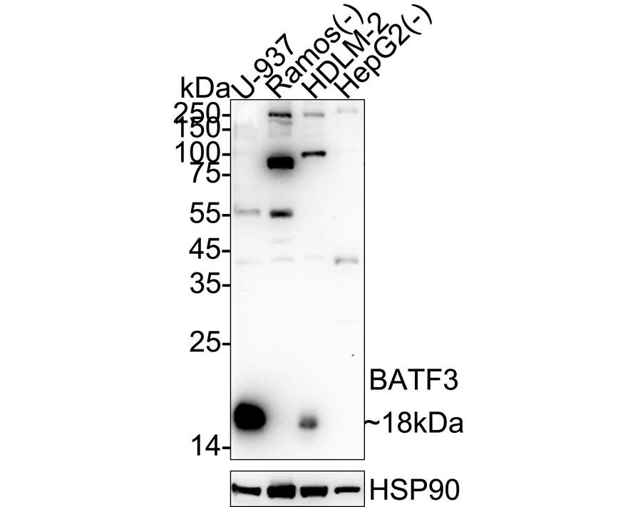
Relative expression (RE). Western blot analysis of BATF3 on different lysates with Rabbit anti-BATF3 antibody at 1/5,000 dilution. Lysates/proteins at 20 µg/Lane. Exposure time: 3 minutes; 4-20% SDS-PAGE gel. Proteins were transferred to a PVDF membrane and blocked with 5% NFDM/TBST for 1 hour at room temperature. The primary antibody at 1/5,000 dilution was used in primary antibody dilution at 4℃ overnight. Goat Anti-Rabbit IgG - HRP Secondary Antibody at 1/50,000 dilution was used for 1 hour at room temperature.IP
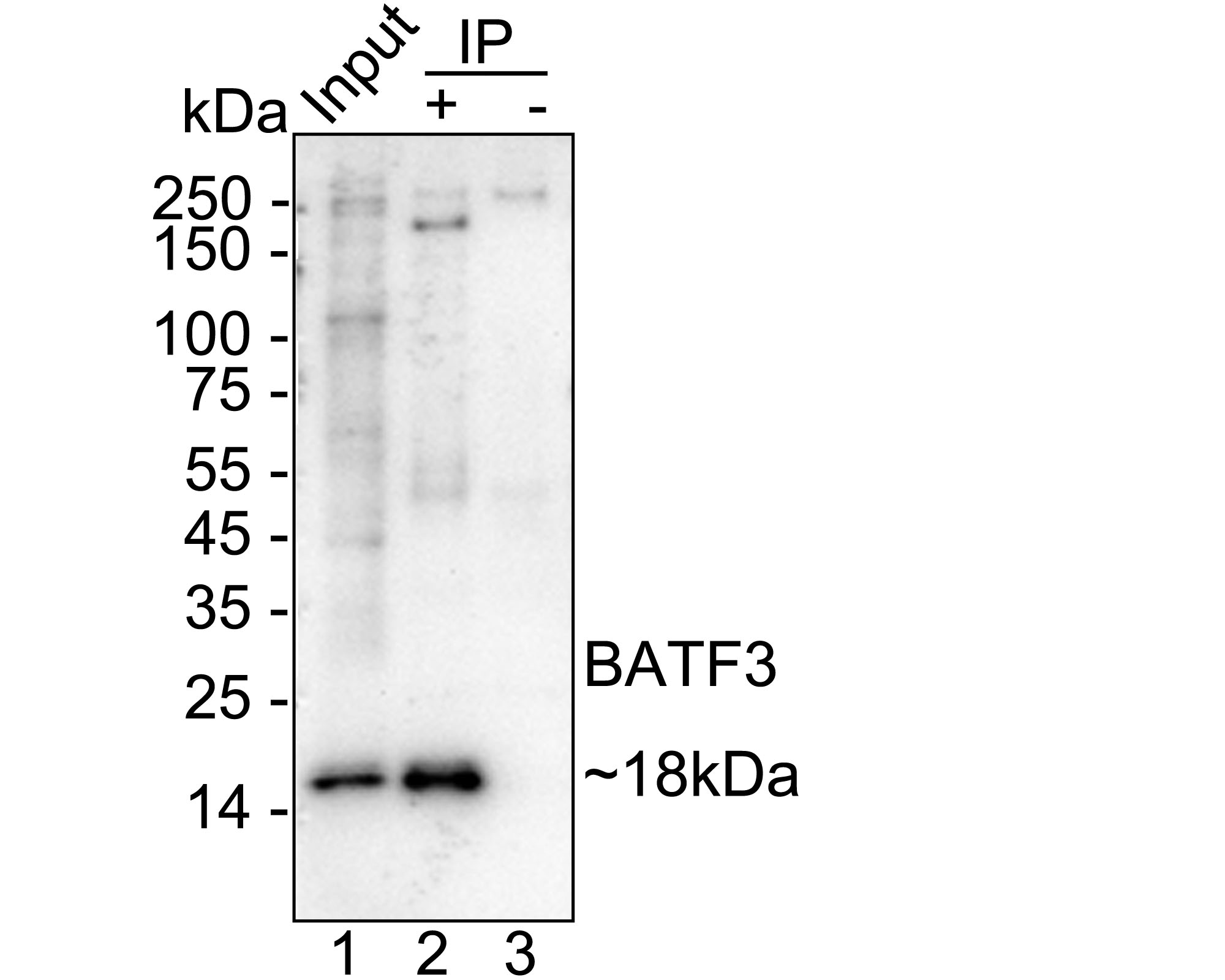
BATF3 was immunoprecipitated from 0.2 mg U-937 cell lysate with BATF3 Recombinant Rabbit Monoclonal Antibody at 2 µg/10 µl beads. Western blot was performed from the immunoprecipitate using BATF3 Recombinant Rabbit Monoclonal Antibody at 1/5,000 dilution. HRP Conjugated Anti-Rabbit IgG for IP Nano-secondary antibody at 1/5,000 dilution was used for 1 hour at room temperature. Lane 1: U-937 cell lysate (input). Lane 2: BATF3 Recombinant Rabbit Monoclonal Antibody IP in U-937 cell lysate. Lane 3: Rabbit IgG instead of BATF3 Recombinant Rabbit Monoclonal Antibody in U-937 cell lysate. Blocking/Dilution buffer: primary antibody dilution . Exposure time: 3 minutes.ChIP
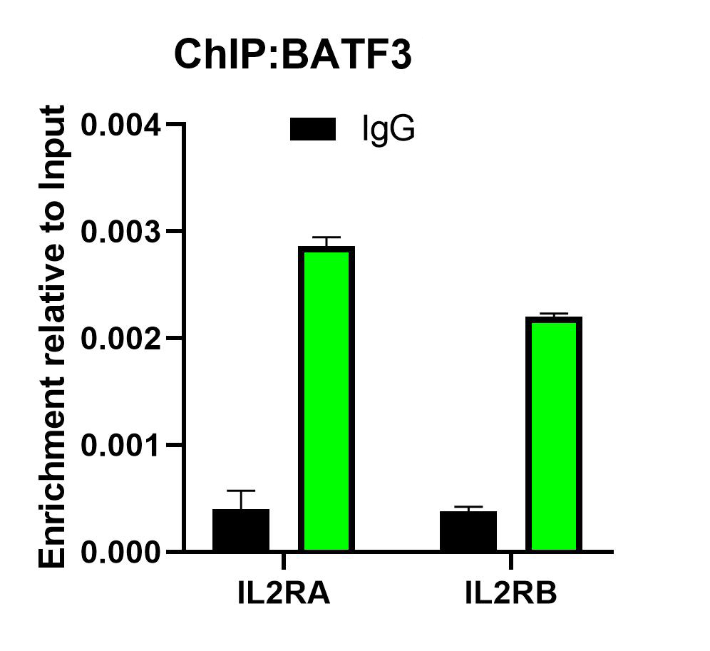
Chromatin immunoprecipitations were performed with cross-linked chromatin from U-937 cells with BATF3 or Normal Rabbit IgG according to the ChIP protocol. The enriched DNA was quantified by real-time PCR using indicated primers. The amount of immunoprecipitated DNA in each sample is represented as signal relative to the total amount of input chromatin, which is equivalent to one.| Product Name | BATF3 Recombinant Rabbit Monoclonal Antibody |
|---|---|
| Antibody Type | Primary Antibodies |
| Immunogen | Recombinant protein within human BATF3 aa 1-127. |
| Clonality | Monoclonal |
|---|---|
| Isotype | IgG |
| Host Species | Rabbit |
| Tested Applications | ChIPIPWB |
| WB:1:5000 IP:1-2μg/sample ChIP:Use 0.5~2 μg for 25 μg of chromatin. |
|
| Species Reactivity | Human |
| Concentration | 1mg/ml |
| Purification | Protein A |
| Gene Symbol | BATF3 |
|---|---|
| Gene Synonyms | JDP1 SNFT JUNDM1 |
| Gene Full Name | basic leucine zipper ATF-like transcription factor 3 |
| Gene Summary | This gene encodes a member of the basic leucine zipper protein family. The encoded protein functions as a transcriptional repressor when heterodimerizing with JUN. The protein may play a role in repression of interleukin-2 and matrix metalloproteinase-1 transcription.[provided by RefSeq, Feb 2009] |
| Alternative Names | 21 kD small nuclear factor isolated from T cells antibody 21 kDa small nuclear factor isolated from T-cells antibody ATF like 3 antibody B-ATF-3 antibody basic leucine zipper transcription factor ATF-like 3 antibody BATF3 antibody FLJ36352 antibody FLJ37535 antibody hCG 39202 antibody JDP1 antibody Jun dimerization protein 1 antibody Jun dimerization protein p21SNFT antibody JUNDM1 antibody SNFT antibody |
| Molecular Weight(MW) | 14 kDa(Observed band size:18 kDa) |
| Cellular Localization | Nucleus. |
WB
Relative expression (RE). Western blot analysis of BATF3 on different lysates with Rabbit anti-BATF3 antibody at 1/5,000 dilution. Lysates/proteins at 20 µg/Lane. Exposure time: 3 minutes; 4-20% SDS-PAGE gel. Proteins were transferred to a PVDF membrane and blocked with 5% NFDM/TBST for 1 hour at room temperature. The primary antibody at 1/5,000 dilution was used in primary antibody dilution at 4℃ overnight. Goat Anti-Rabbit IgG - HRP Secondary Antibody at 1/50,000 dilution was used for 1 hour at room temperature.
IP
BATF3 was immunoprecipitated from 0.2 mg U-937 cell lysate with BATF3 Recombinant Rabbit Monoclonal Antibody at 2 µg/10 µl beads. Western blot was performed from the immunoprecipitate using BATF3 Recombinant Rabbit Monoclonal Antibody at 1/5,000 dilution. HRP Conjugated Anti-Rabbit IgG for IP Nano-secondary antibody at 1/5,000 dilution was used for 1 hour at room temperature. Lane 1: U-937 cell lysate (input). Lane 2: BATF3 Recombinant Rabbit Monoclonal Antibody IP in U-937 cell lysate. Lane 3: Rabbit IgG instead of BATF3 Recombinant Rabbit Monoclonal Antibody in U-937 cell lysate. Blocking/Dilution buffer: primary antibody dilution . Exposure time: 3 minutes.
ChIP
Chromatin immunoprecipitations were performed with cross-linked chromatin from U-937 cells with BATF3 or Normal Rabbit IgG according to the ChIP protocol. The enriched DNA was quantified by real-time PCR using indicated primers. The amount of immunoprecipitated DNA in each sample is represented as signal relative to the total amount of input chromatin, which is equivalent to one.| Application Notes | WB:1:5000 IP:1-2μg/sample ChIP:Use 0.5~2 μg for 25 μg of chromatin. |
|---|
| Form | Liquid |
|---|---|
| Storage Instructions | Shipped at 4℃. Store at +4℃ short term (1-2 weeks). Store at -20℃ long term. |
| Storage Buffer | 1*PBS (pH7.4), 0.1% BSA, 40% Glycerol, 0.2% Proclean 950. |


