WB
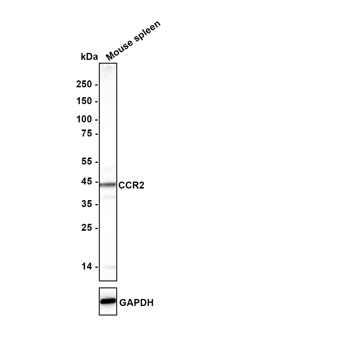
Western blot analysis of CCR2 on different lysates with Rabbit anti-CCR2 antibody at 1/5,000 dilution. Lysates/proteins at 10 µg/Lane. Exposure time: 59 seconds; Blocking: 5% NFDM/TBST, 1 hour at room temperature Primary antibody: CCR2 Recombinant Rabbit Monoclonal Antibody, 1/5,000 in primary antibody dilution buffer, 2 hours at room temperature. Secondary antibody: Goat anti-Rabbit IgG-HRP, 1/50,000 in 5% NFDM/TBST, 1 hour at room temperature.IHC
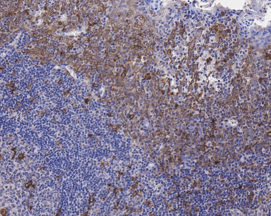
Immunohistochemical analysis of paraffin-embedded human tonsil tissue with Rabbit anti-CCR2 antibody at 1/500 dilution. The section was pre-treated using heat mediated antigen retrieval with Tris-EDTA buffer (pH 9.0) for 20 minutes. The tissues were blocked in 1% BSA for 20 minutes at room temperature, washed with ddH2O and PBS, and then probed with the primary antibody at 1/500 dilution for 1 hour at room temperature. The detection was performed using an HRP conjugated compact polymer system. DAB was used as the chromogen. Tissues were counterstained with hematoxylin and mounted with DPX.FC
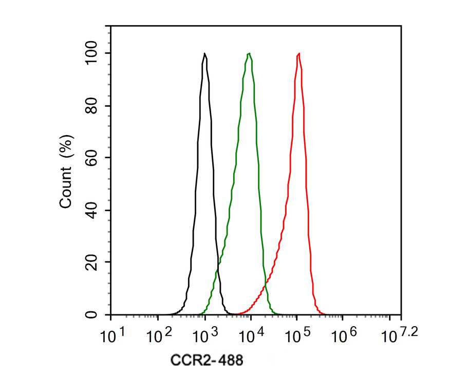
Flow cytometric analysis of THP-1 cells labeling CCR2. Cells were washed twice with cold PBS and resuspend. Then stained with the primary antibody (1ug/ml) (red) compared with Rabbit IgG Isotype Control (green). After incubation of the primary antibody at +4℃ for an hour, the cells were stained with a 488 conjugate-Goat anti-Rabbit IgG Secondary antibody at 1/1,000 dilution for 30 minutes at +4℃. Unlabelled sample was used as a control (cells without incubation with primary antibody; black).| Product Name | CCR2 Recombinant Rabbit Monoclonal Antibody |
|---|---|
| Antibody Type | Primary Antibodies |
| Immunogen | Synthetic peptide within N-terminal human CCR2. |
| Clonality | Monoclonal |
|---|---|
| Isotype | IgG |
| Host Species | Rabbit |
| Tested Applications | FCIHCWB |
| WB:1:2000-1:5000 IHC:1:500 FC:1:500-1:1000 |
|
| Species Reactivity | HumanMouse |
| Concentration | 1mg/ml |
| Purification | Protein A |
| Gene Symbol | CCR2 |
|---|---|
| Gene Synonyms | CKR2 CCR-2 CCR2A CCR2B CD192 CKR2A CKR2B PCLUD CMKBR2 MCP-1-R CC-CKR-2 |
| Gene Full Name | C-C motif chemokine receptor 2 |
| Gene Summary | The protein encoded by this gene is a receptor for monocyte chemoattractant protein-1, a chemokine which specifically mediates monocyte chemotaxis. Monocyte chemoattractant protein-1 is involved in monocyte infiltration in inflammatory diseases such as rheumatoid arthritis as well as in the inflammatory response against tumors. The encoded protein mediates agonist-dependent calcium mobilization and inhibition of adenylyl cyclase. This protein can also be a coreceptor with CD4 for HIV-1 infection. This gene is located in the chemokine receptor gene cluster region of chromosome 3. [provided by RefSeq, Aug 2017] |
| Alternative Names | C C chemokine receptor type 2 antibody C C CKR 2 antibody C-C chemokine receptor type 2 antibody C-C CKR-2 antibody CC chemokine receptor type 2 antibody CC CKR 2 antibody CC-CKR-2 antibody CCCKR2 antibody CCR 2 antibody CCR-2 antibody CCR1L antibody CCR2 antibody CCR2_HUMAN antibody CCR2A antibody CCR2B antibody CCR5L antibody CD192 antibody CD192 antigen antibody Chemokine C C motif receptor 2 antibody Chemokine CC Motif Receptor 2 antibody CKR 2 antibody CKR2 antibody CKR2A antibody CKR2B antibody CMKBR2 antibody MCP 1 R antibody MCP-1-R antibody MCP1 RECEPTOR antibody MCP1R antibody Monocyte chemoattractant protein 1 receptor antibody Monocyte Chemotactic Protein 1 Receptor antibody Monocyte Chemotactic Protein 1 Receptor antibody |
| Molecular Weight(MW) | 42 kDa |
| Cellular Localization | Cell membrane. |
WB
Western blot analysis of CCR2 on different lysates with Rabbit anti-CCR2 antibody at 1/5,000 dilution. Lysates/proteins at 10 µg/Lane. Exposure time: 59 seconds; Blocking: 5% NFDM/TBST, 1 hour at room temperature Primary antibody: CCR2 Recombinant Rabbit Monoclonal Antibody, 1/5,000 in primary antibody dilution buffer, 2 hours at room temperature. Secondary antibody: Goat anti-Rabbit IgG-HRP, 1/50,000 in 5% NFDM/TBST, 1 hour at room temperature.
IHC
Immunohistochemical analysis of paraffin-embedded human tonsil tissue with Rabbit anti-CCR2 antibody at 1/500 dilution. The section was pre-treated using heat mediated antigen retrieval with Tris-EDTA buffer (pH 9.0) for 20 minutes. The tissues were blocked in 1% BSA for 20 minutes at room temperature, washed with ddH2O and PBS, and then probed with the primary antibody at 1/500 dilution for 1 hour at room temperature. The detection was performed using an HRP conjugated compact polymer system. DAB was used as the chromogen. Tissues were counterstained with hematoxylin and mounted with DPX.
FC
Flow cytometric analysis of THP-1 cells labeling CCR2. Cells were washed twice with cold PBS and resuspend. Then stained with the primary antibody (1ug/ml) (red) compared with Rabbit IgG Isotype Control (green). After incubation of the primary antibody at +4℃ for an hour, the cells were stained with a 488 conjugate-Goat anti-Rabbit IgG Secondary antibody at 1/1,000 dilution for 30 minutes at +4℃. Unlabelled sample was used as a control (cells without incubation with primary antibody; black).| Application Notes | WB:1:2000-1:5000 IHC:1:500 FC:1:500-1:1000 |
|---|
| Form | Liquid |
|---|---|
| Storage Instructions | Shipped at 4℃. Store at +4℃ short term (1-2 weeks). Store at -20℃ long term. |
| Storage Buffer | 1*PBS (pH7.4), 0.1% BSA, 40% Glycerol, 0.2% Proclean 950. |


