WB
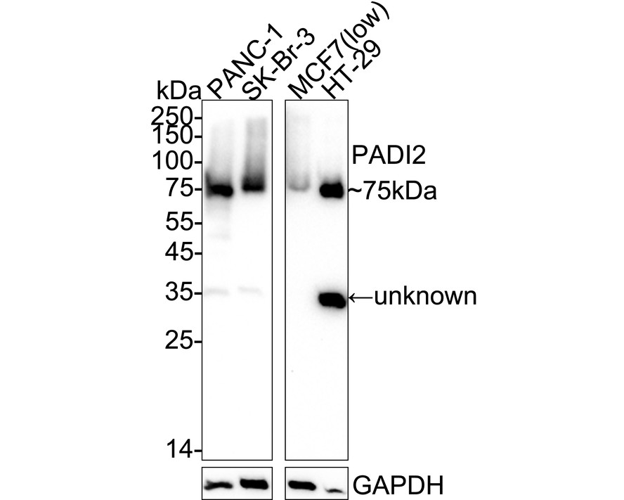
Western blot analysis of PADI2 on different lysates with Rabbit anti-PADI2 antibody at 1/5,000 dilution. Exposure time: Lane 1-2: 6 seconds; Lane 3-5: 3 minutes; 4-20% SDS-PAGE gel. Proteins were transferred to a PVDF membrane and blocked with 5% NFDM/TBST for 1 hour at room temperature. The primary antibody at 1/5,000 dilution was used in primary antibody dilution at 4℃ overnight. Goat Anti-Rabbit IgG - HRP Secondary Antibody at 1/50,000 dilution was used for 1 hour at room temperature.IHC
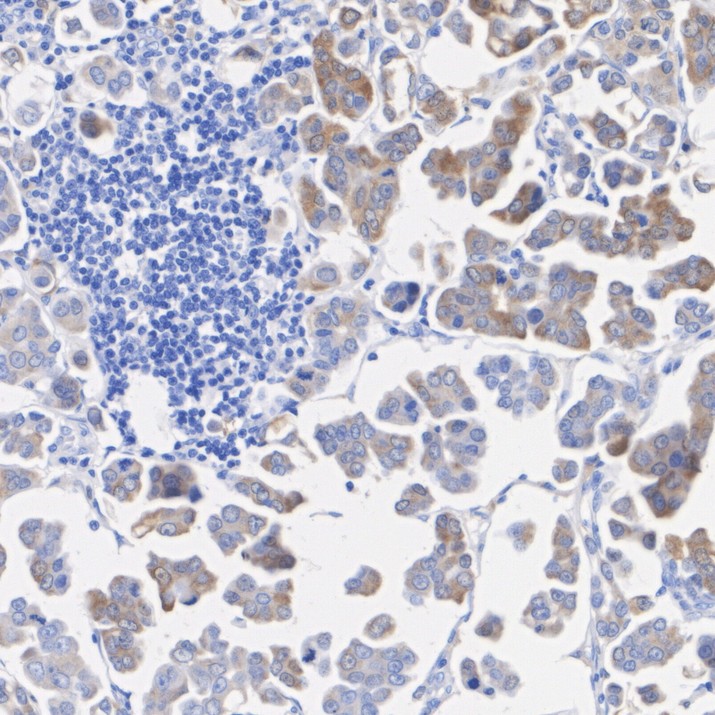
Immunohistochemical analysis of paraffin-embedded human breast cancer tissue with Rabbit anti-PADI2 antibody at 1/200 dilution. The section was pre-treated using heat mediated antigen retrieval with Tris-EDTA buffer (pH 9.0) for 20 minutes. The tissues were blocked in 1% BSA for 20 minutes at room temperature, washed with ddH2O and PBS, and then probed with the primary antibody at 1/200 dilution for 1 hour at room temperature. The detection was performed using an HRP conjugated compact polymer system. DAB was used as the chromogen. Tissues were counterstained with hematoxylin and mounted with DPX.IP
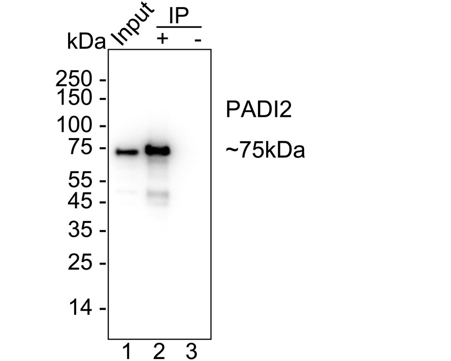
PADI2 was immunoprecipitated from 0.2 mg PANC-1 cell lysate with PADI2 Recombinant Rabbit Monoclonal Antibody at 2 µg/10 µl beads. Western blot was performed from the immunoprecipitate using PADI2 Recombinant Rabbit Monoclonal Antibody at 1/5,000 dilution. HRP Conjugated Anti-Rabbit IgG for IP Nano-secondary antibody at 1/5,000 dilution was used for 1 hour at room temperature. Lane 1: PANC-1 cell lysate (input) Lane 2: PADI2 Recombinant Rabbit Monoclonal Antibody IP in PANC-1 cell lysate Lane 3: Rabbit IgG instead of PADI2 Recombinant Rabbit Monoclonal Antibody in PANC-1 cell lysate Blocking/Dilution buffer: primary antibody dilution. Exposure time: 2 seconds.| Product Name | PADI2 Recombinant Rabbit Monoclonal Antibody |
|---|---|
| Antibody Type | Primary Antibodies |
| Immunogen | Recombinant protein within human PADI2 aa 1-350. |
| Clonality | Monoclonal |
|---|---|
| Isotype | IgG |
| Host Species | Rabbit |
| Tested Applications | IHCIPWB |
| WB:1:5000 IHC:1:200-1:1000 IP:1-2μg/sample |
|
| Species Reactivity | Human |
| Concentration | 1mg/ml |
| Purification | Protein A |
| Gene Symbol | PADI2 |
|---|---|
| Gene Synonyms | PAD2 PDI2 PAD-H19 |
| Gene Full Name | peptidyl arginine deiminase 2 |
| Gene Summary | This gene encodes a member of the peptidyl arginine deiminase family of enzymes, which catalyze the post-translational deimination of proteins by converting arginine residues into citrullines in the presence of calcium ions. The family members have distinct substrate specificities and tissue-specific expression patterns. The type II enzyme is the most widely expressed family member. Known substrates for this enzyme include myelin basic protein in the central nervous system and vimentin in skeletal muscle and macrophages. This enzyme is thought to play a role in the onset and progression of neurodegenerative human disorders, including Alzheimer disease and multiple sclerosis, and it has also been implicated in glaucoma pathogenesis. This gene exists in a cluster with four other paralogous genes. [provided by RefSeq, Jul 2008] |
| Alternative Names | KIAA0994 antibody OTTHUMP00000044625 antibody PAD 2 antibody PAD H19 antibody PAD-H19 antibody PAD2 antibody PADI 2 antibody Padi2 antibody PADI2 protein antibody PADI2_HUMAN antibody PDI 2 antibody PDI2 antibody Peptidlyarginine deiminase type II antibody Peptidyl arginine deiminase II antibody Peptidyl arginine deiminase type II antibody Peptidylarginine deiminase II antibody Protein arginine deiminase antibody Protein arginine deiminase type 2 antibody Protein arginine deiminase type II antibody Protein-arginine deiminase type II antibody Protein-arginine deiminase type-2 antibody |
| Molecular Weight(MW) | 76 kDa(Observed band size:75 kDa) |
| Cellular Localization | Cytoplasm. |
WB
Western blot analysis of PADI2 on different lysates with Rabbit anti-PADI2 antibody at 1/5,000 dilution. Exposure time: Lane 1-2: 6 seconds; Lane 3-5: 3 minutes; 4-20% SDS-PAGE gel. Proteins were transferred to a PVDF membrane and blocked with 5% NFDM/TBST for 1 hour at room temperature. The primary antibody at 1/5,000 dilution was used in primary antibody dilution at 4℃ overnight. Goat Anti-Rabbit IgG - HRP Secondary Antibody at 1/50,000 dilution was used for 1 hour at room temperature.
IHC
Immunohistochemical analysis of paraffin-embedded human breast cancer tissue with Rabbit anti-PADI2 antibody at 1/200 dilution. The section was pre-treated using heat mediated antigen retrieval with Tris-EDTA buffer (pH 9.0) for 20 minutes. The tissues were blocked in 1% BSA for 20 minutes at room temperature, washed with ddH2O and PBS, and then probed with the primary antibody at 1/200 dilution for 1 hour at room temperature. The detection was performed using an HRP conjugated compact polymer system. DAB was used as the chromogen. Tissues were counterstained with hematoxylin and mounted with DPX.
IP
PADI2 was immunoprecipitated from 0.2 mg PANC-1 cell lysate with PADI2 Recombinant Rabbit Monoclonal Antibody at 2 µg/10 µl beads. Western blot was performed from the immunoprecipitate using PADI2 Recombinant Rabbit Monoclonal Antibody at 1/5,000 dilution. HRP Conjugated Anti-Rabbit IgG for IP Nano-secondary antibody at 1/5,000 dilution was used for 1 hour at room temperature. Lane 1: PANC-1 cell lysate (input) Lane 2: PADI2 Recombinant Rabbit Monoclonal Antibody IP in PANC-1 cell lysate Lane 3: Rabbit IgG instead of PADI2 Recombinant Rabbit Monoclonal Antibody in PANC-1 cell lysate Blocking/Dilution buffer: primary antibody dilution. Exposure time: 2 seconds.| Application Notes | WB:1:5000 IHC:1:200-1:1000 IP:1-2μg/sample |
|---|
| Form | Liquid |
|---|---|
| Storage Instructions | Shipped at 4℃. Store at +4℃ short term (1-2 weeks). Store at -20℃ long term. |
| Storage Buffer | 1*PBS (pH7.4), 0.1% BSA, 40% Glycerol, 0.2% Proclean 950. |


