WB
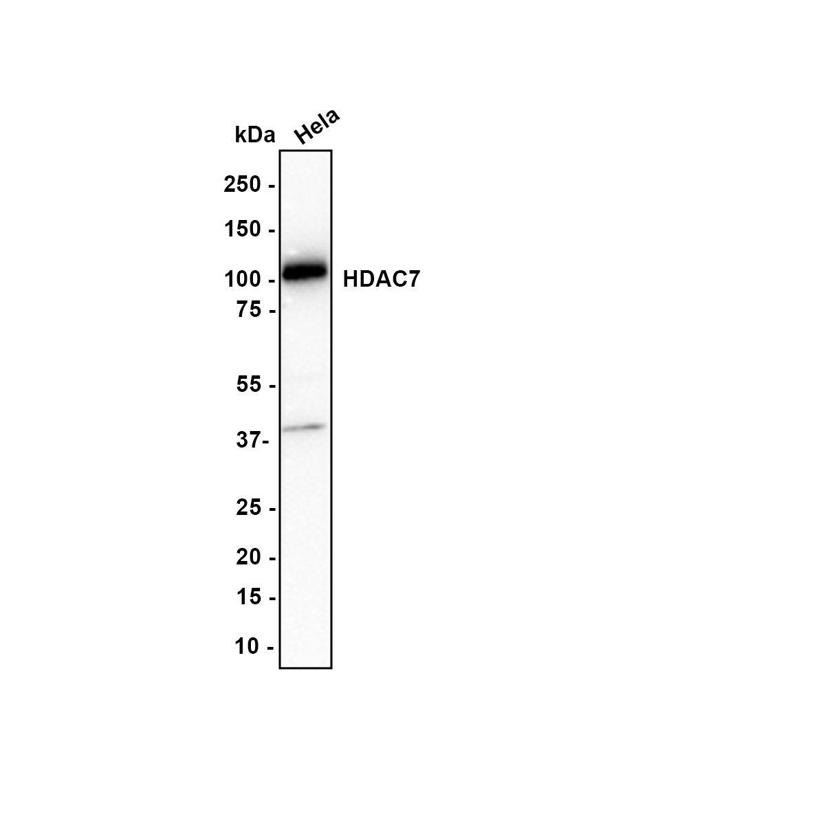
Western blot analysis of HDAC7 on Hela lysates with Rabbit anti-HDAC7 antibody at 1/1,000 dilution. Lysates/proteins at 15 µg/Lane. Exposure time: 3 minutes; Blocking: 5% NFDM/TBST, 1 hour at room temperature Primary antibody: HDAC7 Recombinant Rabbit Monoclonal Antibody, 1/1,000 in primary antibody dilution buffer, overnight at 4 ℃ Secondary antibody: Goat anti-Rabbit IgG-HRP, 1/50,000 in 5% NFDM/TBST, 1 hour at room temperature.ICC/IF
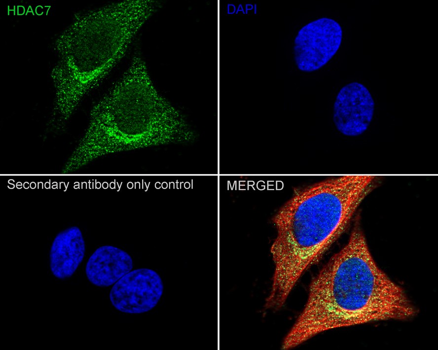
Immunocytochemistry analysis of HeLa cells labeling HDAC7 with Rabbit anti-HDAC7 antibody at 1/100 dilution. Cells were fixed in 4% paraformaldehyde for 15 minutes at room temperature, permeabilized with 0.1% Triton X-100 in PBS for 15 minutes at room temperature, then blocked with 1% BSA in 10% negative goat serum for 1 hour at room temperature. Cells were then incubated with Rabbit anti-HDAC7 antibody at 1/100 dilution in 1% BSA in PBST overnight at 4 ℃. Goat Anti-Rabbit IgG H&L (488) was used as the secondary antibody at 1/1,000 dilution. PBS instead of the primary antibody was used as the secondary antibody only control. Nuclear DNA was labelled in blue with DAPI. Beta tubulin (red) was stained at 1/100 dilution overnight at +4℃. Goat Anti-Mouse IgG H&L (594) was used as the secondary antibody at 1/1,000 dilution.| Product Name | HDAC7 Recombinant Rabbit Monoclonal Antibody |
|---|---|
| Antibody Type | Primary Antibodies |
| Immunogen | Recombinant protein within Human HDAC7 aa 1-218 / 952. |
| Clonality | Monoclonal |
|---|---|
| Isotype | IgG |
| Host Species | Rabbit |
| Tested Applications | ICC/IFWB |
| WB:1:1000-1:2000 ICC/IF:1:100 |
|
| Species Reactivity | HumanMouseRat |
| Concentration | 1mg/ml |
| Purification | Protein A |
| Gene Symbol | HDAC7 |
|---|---|
| Gene Synonyms | HD7 HD7A HDAC7A |
| Gene Full Name | histone deacetylase 7 |
| Gene Summary | Histones play a critical role in transcriptional regulation, cell cycle progression, and developmental events. Histone acetylation/deacetylation alters chromosome structure and affects transcription factor access to DNA. The protein encoded by this gene has sequence homology to members of the histone deacetylase family. This gene is orthologous to mouse HDAC7 gene whose protein promotes repression mediated via the transcriptional corepressor SMRT. Alternatively spliced transcript variants encoding different isoforms have been found for this gene. [provided by RefSeq, Jul 2008] |
| Alternative Names | DKFZP586J0917 antibody FLJ99588 antibody HD 7a antibody HD7 antibody HD7a antibody HDAC 7 antibody HDAC 7A antibody Hdac7 antibody HDAC7_HUMAN antibody HDAC7A antibody Histone deacetylase 7 antibody Histone deacetylase 7A antibody OTTHUMP00000202813 antibody OTTHUMP00000202814 antibody |
| Molecular Weight(MW) | 103 kDa |
| Cellular Localization | Nucleus, Cytoplasm. |
WB
Western blot analysis of HDAC7 on Hela lysates with Rabbit anti-HDAC7 antibody at 1/1,000 dilution. Lysates/proteins at 15 µg/Lane. Exposure time: 3 minutes; Blocking: 5% NFDM/TBST, 1 hour at room temperature Primary antibody: HDAC7 Recombinant Rabbit Monoclonal Antibody, 1/1,000 in primary antibody dilution buffer, overnight at 4 ℃ Secondary antibody: Goat anti-Rabbit IgG-HRP, 1/50,000 in 5% NFDM/TBST, 1 hour at room temperature.
ICC/IF
Immunocytochemistry analysis of HeLa cells labeling HDAC7 with Rabbit anti-HDAC7 antibody at 1/100 dilution. Cells were fixed in 4% paraformaldehyde for 15 minutes at room temperature, permeabilized with 0.1% Triton X-100 in PBS for 15 minutes at room temperature, then blocked with 1% BSA in 10% negative goat serum for 1 hour at room temperature. Cells were then incubated with Rabbit anti-HDAC7 antibody at 1/100 dilution in 1% BSA in PBST overnight at 4 ℃. Goat Anti-Rabbit IgG H&L (488) was used as the secondary antibody at 1/1,000 dilution. PBS instead of the primary antibody was used as the secondary antibody only control. Nuclear DNA was labelled in blue with DAPI. Beta tubulin (red) was stained at 1/100 dilution overnight at +4℃. Goat Anti-Mouse IgG H&L (594) was used as the secondary antibody at 1/1,000 dilution.| Application Notes | WB:1:1000-1:2000 ICC/IF:1:100 |
|---|
| Form | Liquid |
|---|---|
| Storage Instructions | Shipped at 4℃. Store at +4℃ short term (1-2 weeks). Store at -20℃ long term. |
| Storage Buffer | 1*TBS (pH7.4), 0.05% BSA, 40% Glycerol. Preservative: 0.05% Sodium Azide. |


