WB
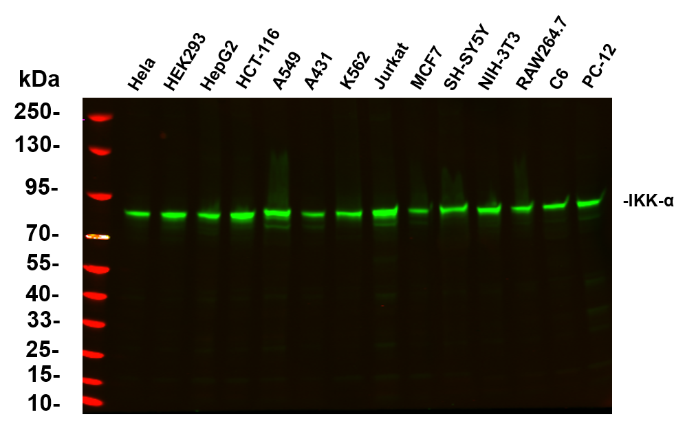
Various whole cell lysates were separated by 4-20% SDS-PAGE, and the primary antibody was used at 4℃ over night with a 1:5000 dilution . The Dylight 800-conjugated Goat anti-Rabbit antibody was used to detect the antibody.IHC
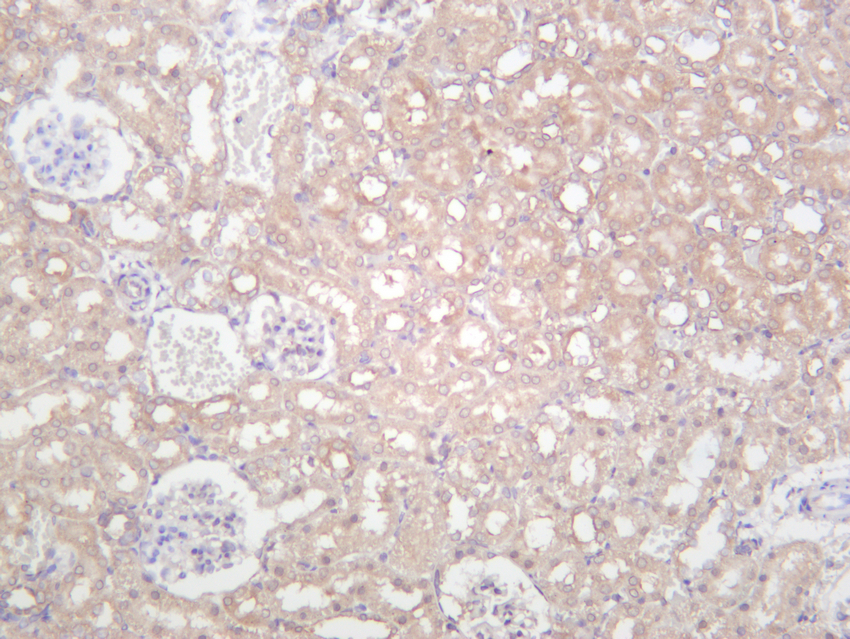
Rat kidney was stained with anti-IKK α rabbit antibody.IHC
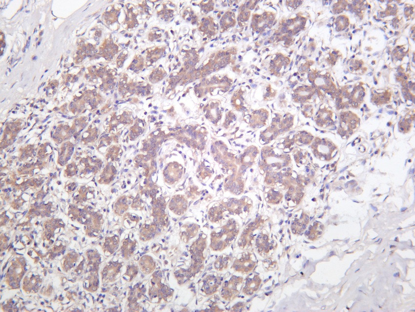
Human breast carcinoma was stained with anti-IKK α rabbit antibody.IHC
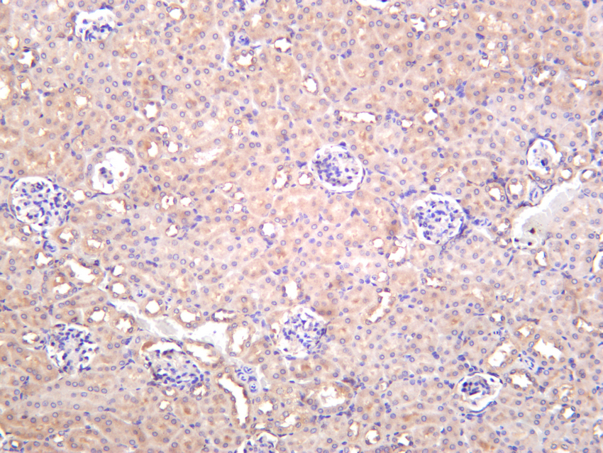
Mouse kidney was stained with anti-IKK α rabbit antibody.ICC/IF
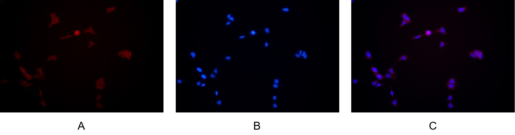
Immunofluorescence analysis of HEK293. Picture A:IKK α antibody (red). Picture B: DAPI (blue). Picture C: Merge of A+B.| Product Name | IKK-α Rabbit mAb |
|---|---|
| Antibody Type | Primary Antibodies |
| Immunogen | The specific immunogen used to produce this antibody is proprietary information. |
| Clonality | Monoclonal |
|---|---|
| Isotype | IgG,K |
| Host Species | Rabbit |
| Tested Applications | ICC/IFIHCWB |
| WB:1:2000-1:10000 IHC:1:100-1:500 ICC/IF:1:200-1:1000 |
|
| Species Reactivity | HumanMouseRat |
| Concentration | 1mg/ml |
| Purification | Protein A |
| Gene Symbol | CHUK |
|---|---|
| Gene Synonyms | BPS2 IKK1 IKKA IKBKA IKK-1 TCF16 NFKBIKA IKK-alpha |
| Gene Full Name | component of inhibitor of nuclear factor kappa B kinase complex |
| Gene Summary | This gene encodes a member of the serine/threonine protein kinase family. The encoded protein, a component of a cytokine-activated protein complex that is an inhibitor of the essential transcription factor NF-kappa-B complex, phosphorylates sites that trigger the degradation of the inhibitor via the ubiquination pathway, thereby activating the transcription factor. [provided by RefSeq, Jul 2008] |
| Alternative Names |
CHUK IKKA TCF16 Inhibitor of nuclear factor kappa-B kinase subunit alpha I-kappa-B kinase alpha IKK-A IKK-alpha IkBKA IkappaB kinase Conserved helix-loop-helix ubiquitous kinase I-kappa-B kinase 1 IKK1 Nuclear factor NF-kappa-B |
| Molecular Weight(MW) | 85 kDa |
| Cellular Localization | Cytoplasm , Nucleus. |
WB
Various whole cell lysates were separated by 4-20% SDS-PAGE, and the primary antibody was used at 4℃ over night with a 1:5000 dilution . The Dylight 800-conjugated Goat anti-Rabbit antibody was used to detect the antibody.
IHC
Rat kidney was stained with anti-IKK α rabbit antibody.
IHC
Human breast carcinoma was stained with anti-IKK α rabbit antibody.
IHC
Mouse kidney was stained with anti-IKK α rabbit antibody.
ICC/IF
Immunofluorescence analysis of HEK293. Picture A:IKK α antibody (red). Picture B: DAPI (blue). Picture C: Merge of A+B.| Application Notes | WB:1:2000-1:10000 IHC:1:100-1:500 ICC/IF:1:200-1:1000 |
|---|
| Form | Liquid |
|---|---|
| Storage Instructions | -15°C to -25°C/1 year (Do not lower than -25°C). |
| Storage Buffer | PBS, 50% glycerol, 0.05% Proclin 300, 0.05%BSA. |


