WB
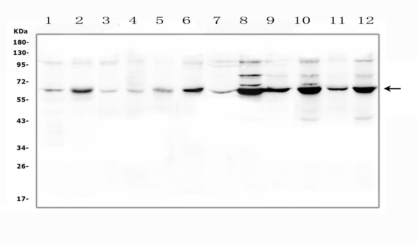
Western blot analysis of SLC19A1 using anti-SLC19A1 antibody. The sample well of each lane was loaded with 30 ug of sample under reducing conditions. Lane 1: rat brain tissue lysates, Lane 2: rat ovary tissue lysates, Lane 3: mouse brain tissue lysates, Lane 4: mouse testicular tissue lysates, Lane 5: mouse ovary tissue lysates, Lane 6: mouse NIH/3T3 whole cell lysates, Lane 7: human placenta tissue lysates, Lane 8: human HEK293 whole cell lysates, Lane 9: monkey COS-7 whole cell lysates, Lane 10: human PC-3 whole cell lysates, Lane 11: human 22RV1 whole cell lysates, Lane 12: human K562 whole cell lysates. After electrophoresis, proteins were transferred to a membrane. Then the membrane was incubated with rabbit anti-SLC19A1 antigen affinity purified polyclonal antibody at a dilution of 1:1000 and probed with a goat anti-rabbit IgG-HRP secondary antibody. The signal is developed using ECL Plus Western Blotting Substrate.| Product Name | Rabbit polyclonal antibody to SLC19A1 |
|---|---|
| Antibody Type | Primary Antibodies |
| Immunogen | A synthetic peptide corresponding to a sequence in the middle region of human SLC19A1. |
| Clonality | Polyclonal |
|---|---|
| Isotype | IgG |
| Host Species | Rabbit |
| Tested Applications | WB |
| WB:1:500-1:2000 |
|
| Species Reactivity | HumanMonkeyMouseRat |
| Concentration | 0.5mg/ml |
| Purification | Affinity purified |
| Gene Symbol | SLC19A1 |
|---|---|
| Gene Synonyms | RFC CHMD FOLT IFC1 REFC RFC1 hRFC IFC-1 MEGAF RFT-1 IMD114 hSLC19A1 |
| Gene Full Name | solute carrier family 19 member 1 |
| Gene Summary | The membrane protein encoded by this gene is a transporter of folate and is involved in the regulation of intracellular concentrations of folate. Three transcript variants encoding different isoforms have been found for this gene.[provided by RefSeq, Mar 2011] |
| Alternative Names | CHMD FLOT1 Folate transporter 1 FOLT IFC 1 IFC1 Intestinal folate carrier 1 Placental folate transporter Reduced folate carrier protein REFC RFC RFC1 SLC19A1 |
| Molecular Weight(MW) | 65 kDa |
| Cellular Localization | Cell membrane,embrane. |
WB
Western blot analysis of SLC19A1 using anti-SLC19A1 antibody. The sample well of each lane was loaded with 30 ug of sample under reducing conditions. Lane 1: rat brain tissue lysates, Lane 2: rat ovary tissue lysates, Lane 3: mouse brain tissue lysates, Lane 4: mouse testicular tissue lysates, Lane 5: mouse ovary tissue lysates, Lane 6: mouse NIH/3T3 whole cell lysates, Lane 7: human placenta tissue lysates, Lane 8: human HEK293 whole cell lysates, Lane 9: monkey COS-7 whole cell lysates, Lane 10: human PC-3 whole cell lysates, Lane 11: human 22RV1 whole cell lysates, Lane 12: human K562 whole cell lysates. After electrophoresis, proteins were transferred to a membrane. Then the membrane was incubated with rabbit anti-SLC19A1 antigen affinity purified polyclonal antibody at a dilution of 1:1000 and probed with a goat anti-rabbit IgG-HRP secondary antibody. The signal is developed using ECL Plus Western Blotting Substrate.| Application Notes | WB:1:500-1:2000 |
|---|
| Form | Liquid |
|---|---|
| Storage Instructions | 12 months from date of receipt,-20℃ as supplied. |
| Storage Buffer | 500 ug/ml antibody with PBS, 0.02% NaN3, 1 mg/ml BSA and 50% glycerol. |


