WB
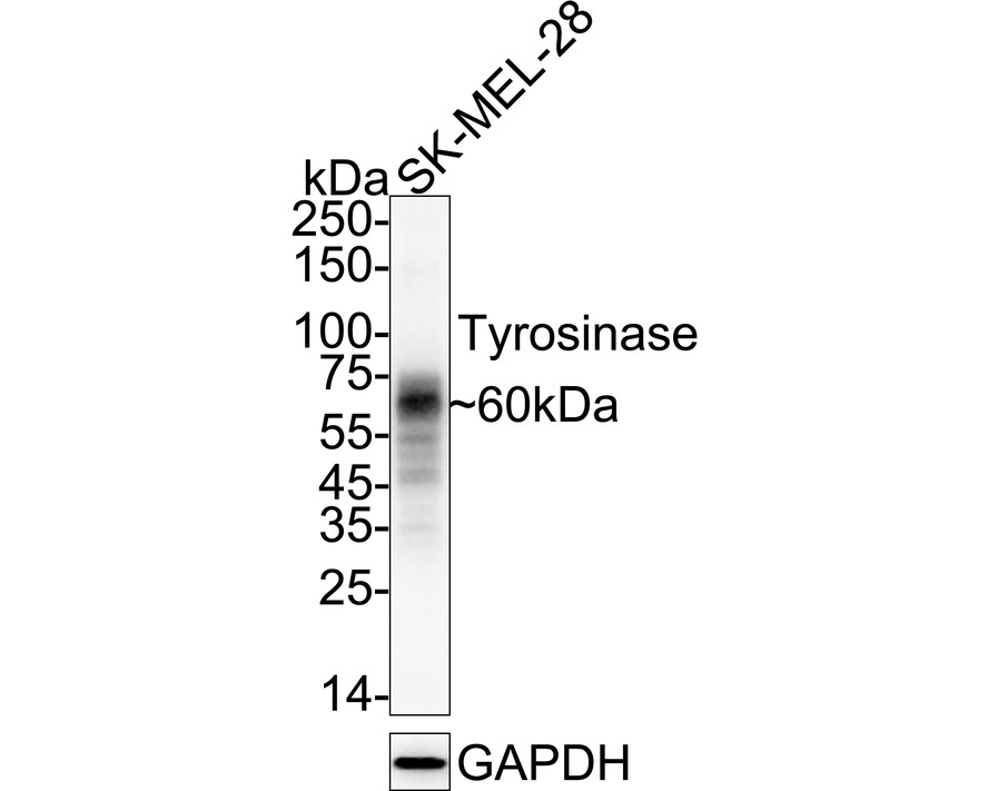
Western blot analysis of Tyrosinase on SK-MEL-28 cell lysates with Rabbit anti-Tyrosinase antibody at 1/2,000 dilution. Lysates/proteins at 20 µg/Lane. Exposure time: 46 seconds; 4-20% SDS-PAGE gel. Proteins were transferred to a PVDF membrane and blocked with 5% NFDM/TBST for 1 hour at room temperature. The primary antibody at 1/2,000 dilution was used in 5% NFDM/TBST at 4℃ overnight. Goat Anti-Rabbit IgG - HRP Secondary Antibody at 1/50,000 dilution was used for 1 hour at room temperature.IHC
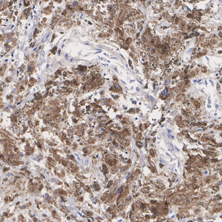
Application: Immunohistochemistry (IHC-P). Species: Human. Tissue: Melanoma. Sample: Paraffin-embedded section. Antigen retrieval: Heat-mediated, Tris-EDTA buffer (pH 9.0), 20 minutes at 95℃. Wash buffer: 1× TBST Endogenous peroxidase blocking: 3% H₂O₂, 10 minutes at room temperature. Blocking: 1% BSA + 10% normal goat serum, 10 minutes at room temperature. Primary antibody: Tyrosinase Recombinant Rabbit Monoclonal Antibody, 1/5,000, 1 hour at room temperature. Secondary antibody: 20 minutes at room temperature.ICC/IF
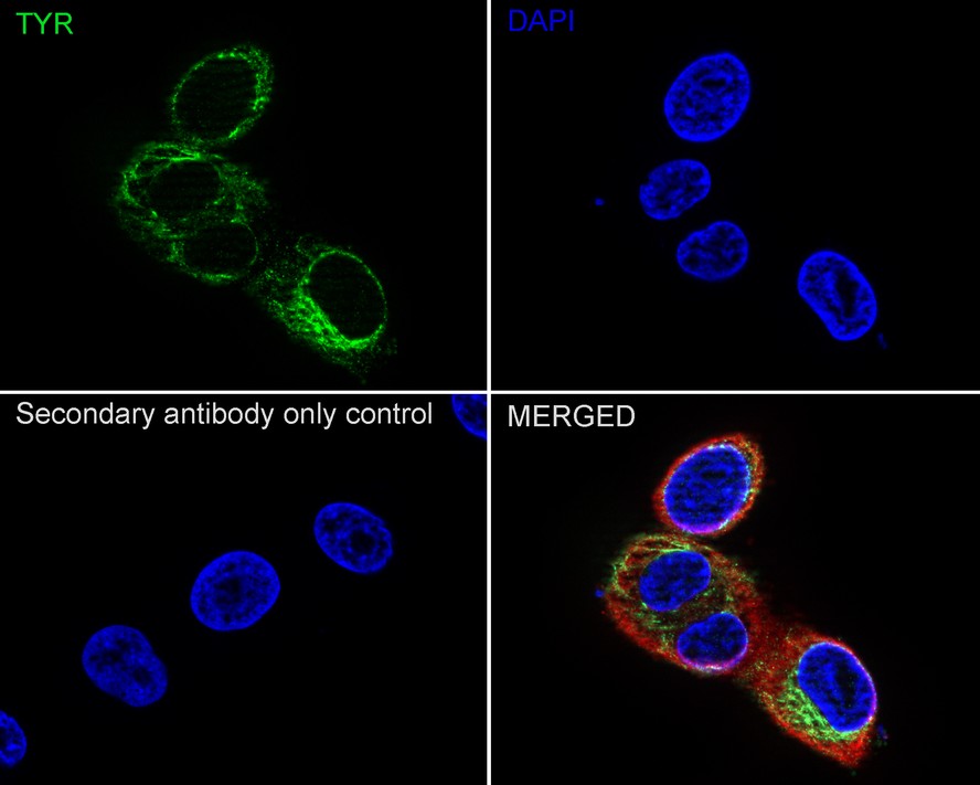
Immunocytochemistry analysis of SK-MEL-28 cells labeling Tyrosinase with Rabbit anti-Tyrosinase antibody at 1/100 dilution. Cells were fixed in 4% paraformaldehyde for 15 minutes at room temperature, permeabilized with 0.1% Triton X-100 in PBS for 15 minutes at room temperature, then blocked with 1% BSA in 10% negative goat serum for 1 hour at room temperature. Cells were then incubated with Rabbit anti-Tyrosinase antibody at 1/100 dilution in 1% BSA in PBST overnight at 4 ℃. Goat Anti-Rabbit IgG H&L (488) was used as the secondary antibody at 1/1,000 dilution. PBS instead of the primary antibody was used as the secondary antibody only control. Nuclear DNA was labelled in blue with DAPI. Beta tubulin (red) was stained at 1/100 dilution overnight at +4℃. Goat Anti-Mouse IgG H&L (594) was used as the secondary antibody at 1/1,000 dilution.FC
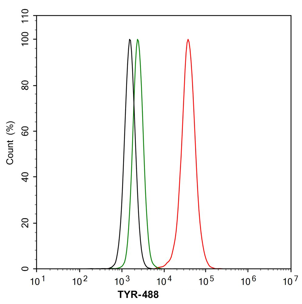
Flow cytometric analysis of SK-MEL-28 cells labeling Tyrosinase. Cells were washed twice with cold PBS and resuspend. Then stained with the primary antibody (1/1,000) (red) compared with Rabbit IgG Isotype Control (green). After incubation of the primary antibody at +4℃ for an hour, the cells were stained with a 488 conjugate-Goat anti-Rabbit IgG Secondary antibody at 1/1,000 dilution for 30 minutes at +4℃. Unlabelled sample was used as a control (cells without incubation with primary antibody; black).IP
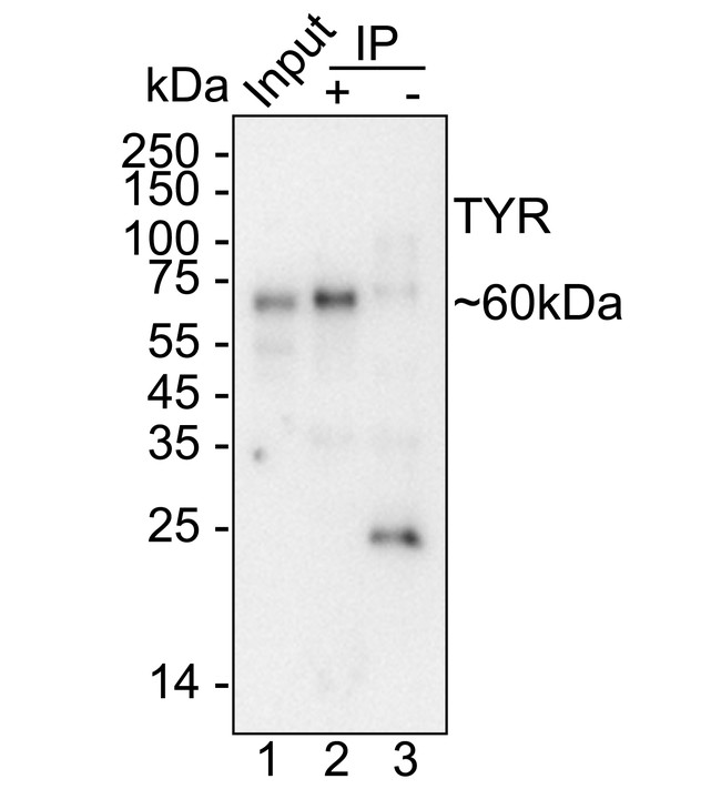
Tyrosinase was immunoprecipitated from 0.2 mg SK-MEL-28 cell lysate with Tyrosinase Recombinant Rabbit Monoclonal Antibody at 2 µg/10 µl beads. Western blot was performed from the immunoprecipitate using Tyrosinase Recombinant Rabbit Monoclonal Antibody at 1/2,000 dilution. HRP Conjugated Anti-Rabbit IgG for IP Nano-secondary antibody at 1/5,000 dilution was used for 1 hour at room temperature. Lane 1: SK-MEL-28 cell lysate (input). Lane 2: Tyrosinase Recombinant Rabbit Monoclonal Antibody IP in SK-MEL-28 cell lysate. Lane 3: Rabbit IgG instead of Tyrosinase Recombinant Rabbit Monoclonal Antibody in SK-MEL-28 cell lysate. Blocking/Dilution buffer: 5% NFDM/TBST. Exposure time: 3 minutes.| Product Name | Tyrosinase Recombinant Rabbit Monoclonal Antibody |
|---|---|
| Antibody Type | Primary Antibodies |
| Immunogen | Recombinant protein within Human Tyrosinase aa 1-340 / 529. |
| Clonality | Monoclonal |
|---|---|
| Isotype | IgG |
| Host Species | Rabbit |
| Tested Applications | FCICC/IFIHCIPWB |
| WB:1:500-1:2000 IHC:1:5000 ICC/IF:1:100 FC:1:1000 IP:1-2μg/sample |
|
| Species Reactivity | HumanMouse |
| Concentration | 1mg/ml |
| Purification | Protein A |
| Gene Symbol | TYR |
|---|---|
| Gene Synonyms | ATN CMM8 OCA1 OCA1A OCAIA SHEP3 |
| Gene Full Name | tyrosinase |
| Gene Summary | The enzyme encoded by this gene catalyzes the first 2 steps, and at least 1 subsequent step, in the conversion of tyrosine to melanin. The enzyme has both tyrosine hydroxylase and dopa oxidase catalytic activities, and requires copper for function. Mutations in this gene result in oculocutaneous albinism, and nonpathologic polymorphisms result in skin pigmentation variation. The human genome contains a pseudogene similar to the 3' half of this gene. [provided by RefSeq, Oct 2008] |
| Alternative Names | ATN antibody CMM8 antibody LB24 AB antibody LB24-AB antibody Monophenol monooxygenase antibody OCA1 antibody OCA1A antibody OCAIA antibody Oculocutaneous albinism IA antibody SHEP3 antibody SK29 AB antibody SK29-AB antibody Tumor rejection antigen AB antibody TYR antibody TYRO_HUMAN antibody tyrosinase (oculocutaneous albinism IA) antibody Tyrosinase antibody |
| Molecular Weight(MW) | 60 kDa |
| Cellular Localization | Melanosome membrane, Melanosome. |
WB
Western blot analysis of Tyrosinase on SK-MEL-28 cell lysates with Rabbit anti-Tyrosinase antibody at 1/2,000 dilution. Lysates/proteins at 20 µg/Lane. Exposure time: 46 seconds; 4-20% SDS-PAGE gel. Proteins were transferred to a PVDF membrane and blocked with 5% NFDM/TBST for 1 hour at room temperature. The primary antibody at 1/2,000 dilution was used in 5% NFDM/TBST at 4℃ overnight. Goat Anti-Rabbit IgG - HRP Secondary Antibody at 1/50,000 dilution was used for 1 hour at room temperature.
IHC
Application: Immunohistochemistry (IHC-P). Species: Human. Tissue: Melanoma. Sample: Paraffin-embedded section. Antigen retrieval: Heat-mediated, Tris-EDTA buffer (pH 9.0), 20 minutes at 95℃. Wash buffer: 1× TBST Endogenous peroxidase blocking: 3% H₂O₂, 10 minutes at room temperature. Blocking: 1% BSA + 10% normal goat serum, 10 minutes at room temperature. Primary antibody: Tyrosinase Recombinant Rabbit Monoclonal Antibody, 1/5,000, 1 hour at room temperature. Secondary antibody: 20 minutes at room temperature.
ICC/IF
Immunocytochemistry analysis of SK-MEL-28 cells labeling Tyrosinase with Rabbit anti-Tyrosinase antibody at 1/100 dilution. Cells were fixed in 4% paraformaldehyde for 15 minutes at room temperature, permeabilized with 0.1% Triton X-100 in PBS for 15 minutes at room temperature, then blocked with 1% BSA in 10% negative goat serum for 1 hour at room temperature. Cells were then incubated with Rabbit anti-Tyrosinase antibody at 1/100 dilution in 1% BSA in PBST overnight at 4 ℃. Goat Anti-Rabbit IgG H&L (488) was used as the secondary antibody at 1/1,000 dilution. PBS instead of the primary antibody was used as the secondary antibody only control. Nuclear DNA was labelled in blue with DAPI. Beta tubulin (red) was stained at 1/100 dilution overnight at +4℃. Goat Anti-Mouse IgG H&L (594) was used as the secondary antibody at 1/1,000 dilution.
FC
Flow cytometric analysis of SK-MEL-28 cells labeling Tyrosinase. Cells were washed twice with cold PBS and resuspend. Then stained with the primary antibody (1/1,000) (red) compared with Rabbit IgG Isotype Control (green). After incubation of the primary antibody at +4℃ for an hour, the cells were stained with a 488 conjugate-Goat anti-Rabbit IgG Secondary antibody at 1/1,000 dilution for 30 minutes at +4℃. Unlabelled sample was used as a control (cells without incubation with primary antibody; black).
IP
Tyrosinase was immunoprecipitated from 0.2 mg SK-MEL-28 cell lysate with Tyrosinase Recombinant Rabbit Monoclonal Antibody at 2 µg/10 µl beads. Western blot was performed from the immunoprecipitate using Tyrosinase Recombinant Rabbit Monoclonal Antibody at 1/2,000 dilution. HRP Conjugated Anti-Rabbit IgG for IP Nano-secondary antibody at 1/5,000 dilution was used for 1 hour at room temperature. Lane 1: SK-MEL-28 cell lysate (input). Lane 2: Tyrosinase Recombinant Rabbit Monoclonal Antibody IP in SK-MEL-28 cell lysate. Lane 3: Rabbit IgG instead of Tyrosinase Recombinant Rabbit Monoclonal Antibody in SK-MEL-28 cell lysate. Blocking/Dilution buffer: 5% NFDM/TBST. Exposure time: 3 minutes.| Application Notes | WB:1:500-1:2000 IHC:1:5000 ICC/IF:1:100 FC:1:1000 IP:1-2μg/sample |
|---|
| Form | Liquid |
|---|---|
| Storage Instructions | Shipped at 4℃. Store at +4℃ short term (1-2 weeks). Store at -20℃ long term. |
| Storage Buffer | 1*PBS (pH7.4), 0.1% BSA, 40% Glycerol, 0.2% Proclean 950. |


