WB
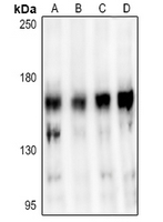
Western blot analysis of Pyruvate Kinase expression in HCT116 (A), COS7 (B), K562 (C), A549 (D), Hela (E) whole cell lysates.ICC/IF
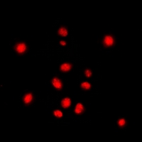
Immunofluorescent analysis of Pyruvate Kinase staining in NIH3T3 cells. Formalin-fixed cells were permeabilized with 0.1% Triton X-100 in TBS for 5-10 minutes and blocked with 3% BSA-PBS for 30 minutes at room temperature. Cells were probed with the primary antibody in 3% BSA-PBS and incubated overnight at 4 °C in a hidified chamber. Cells were washed with PBST and incubated with a AF488-conjugated secondary antibody (green) in PBS at room temperature in the dark.IHC
| Product Name | STAG3 polyclonal antibody |
|---|---|
| Antibody Type | Primary Antibodies |
| Immunogen | A synthetic peptide encompassing a sequence within the C-term region of human STAG3. |
| Clonality | Polyclonal |
|---|---|
| Isotype | IgG |
| Host Species | Rabbit |
| Tested Applications | ICC/IFIHCWB |
| WB:1:500-1:1000 IHC:1:100 - 1:200 ICC/IF:1:100 - 1:500 |
|
| Species Reactivity | HumanMouse |
| Concentration | 1mg/ml |
| Purification | Affinity purified |
| Gene Symbol | STAG3 |
|---|---|
| Gene Synonyms | SA3 SPGF61 |
| Gene Full Name | STAG3 cohesin complex component |
| Gene Summary | The protein encoded by this gene is expressed in the nucleus and is a subunit of the cohesin complex which regulates the cohesion of sister chromatids during cell division. A mutation in this gene is associated with premature ovarian failure. Alternate splicing results in multiple transcript variants encoding distinct isoforms. This gene has multiple pseudogenes. [provided by RefSeq, Apr 2014] |
| Alternative Names | Cohesin subunit SA-3 SCC3 homolog 3 Stromal antigen 3 Stromalin-3 |
| Molecular Weight(MW) | ~139 kDa |
| Cellular Localization | Centromere,Chromosome,Nucleus. |
WB
Western blot analysis of Pyruvate Kinase expression in HCT116 (A), COS7 (B), K562 (C), A549 (D), Hela (E) whole cell lysates.
ICC/IF
Immunofluorescent analysis of Pyruvate Kinase staining in NIH3T3 cells. Formalin-fixed cells were permeabilized with 0.1% Triton X-100 in TBS for 5-10 minutes and blocked with 3% BSA-PBS for 30 minutes at room temperature. Cells were probed with the primary antibody in 3% BSA-PBS and incubated overnight at 4 °C in a hidified chamber. Cells were washed with PBST and incubated with a AF488-conjugated secondary antibody (green) in PBS at room temperature in the dark.
IHC
| Application Notes | WB:1:500-1:1000 IHC:1:100 - 1:200 ICC/IF:1:100 - 1:500 |
|---|
| Form | Liquid |
|---|---|
| Storage Instructions | Store at 4°C short term. Aliquot and store at -20°C long term. Avoid freeze-thaw cycles. |
| Storage Buffer | 1mg/ml in 0.42% Potassium phosphate, 0.87% Sodium chloride, pH 7.3, 30% glycerol, and 0.01% sodium azide. |


