WB
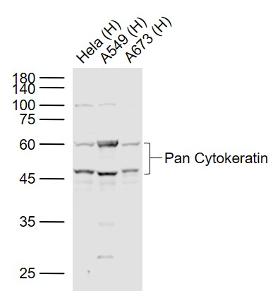
Sample: Lane 1: Hela (Human) Cell Lysate at 30 ug, Lane 2: A549 (Human) Cell Lysate at 30 ug, Lane 3: A673 (Human) Cell Lysate at 30 ug, Primary: Anti-Pan Cytokeratin at 1/1000 dilution, Secondary: IRDye800CW Goat Anti-Rabbit IgG at 1/20000 dilution.IHC
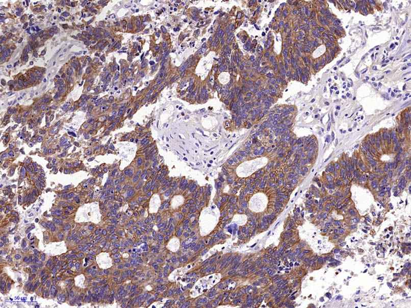
Paraformaldehyde-fixed, paraffin embedded (Human stomach carcinoma); Antigen retrieval by boiling in sodium citrate buffer (pH6.0) for 15min; Block endogenous peroxidase by 3% hydrogen peroxide for 20 minutes; Blocking buffer (normal goat serum) at 37°C for 30min; Antibody incubation with (Pan Cytokeratin) Polyclonal Antibody at 1:400 overnight at 4°C, followed by operating according to SP Kit(Rabbit) instructionsand DAB staining.IHC
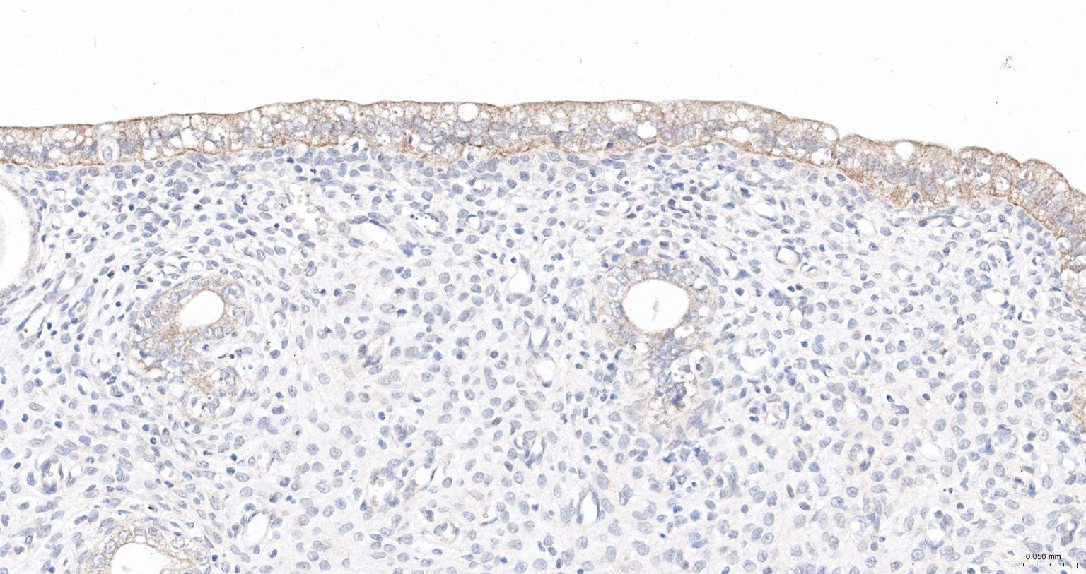
Paraformaldehyde-fixed, paraffin embedded Rat Uterus; Antigen retrieval by boiling in sodium citrate buffer (pH6.0) for 15 min; The section was incubated with Pan Cytokeratin Polyclonal Antibody, Unconjugated at 1:200 overnight at 4°C, followed by conjugation to the Goat anti-Rabbit IgG (H&L) pAb-HRP and DAB staining.ICC/IF
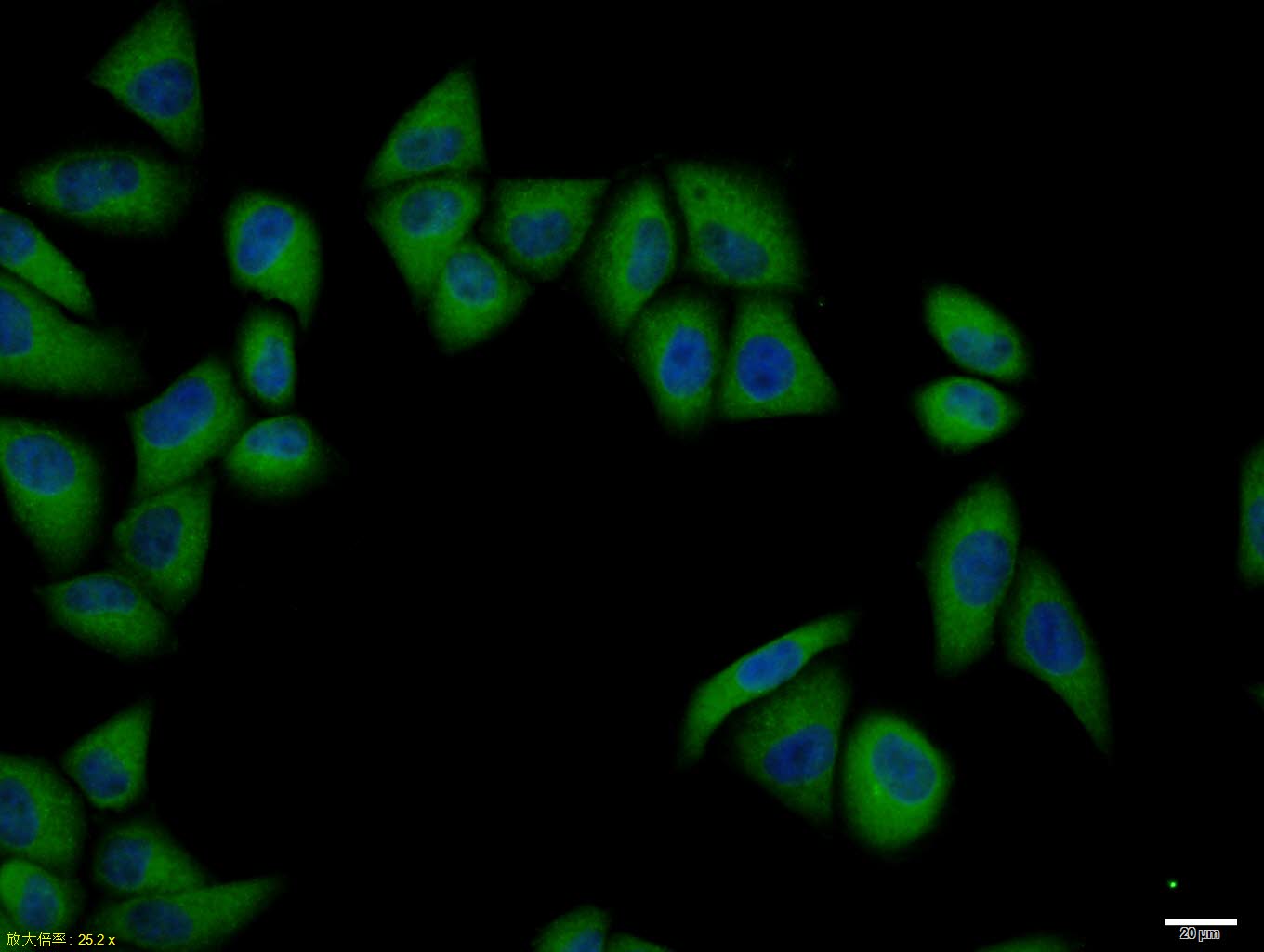
Hela cell; 4% Paraformaldehyde-fixed; Triton X-100 at room temperature for 20 min; Blocking buffer (normal goat serum) at 37°C for 20 min; Antibody incubation with (Pan Cytokeratin) polyclonal Antibody 1:100, 90 minutes at 37°C; followed by a conjugated Goat Anti-Rabbit IgG antibody at 37°C for 90 minutes, DAPI (blue) was used to stain the cell nuclei.IF-P
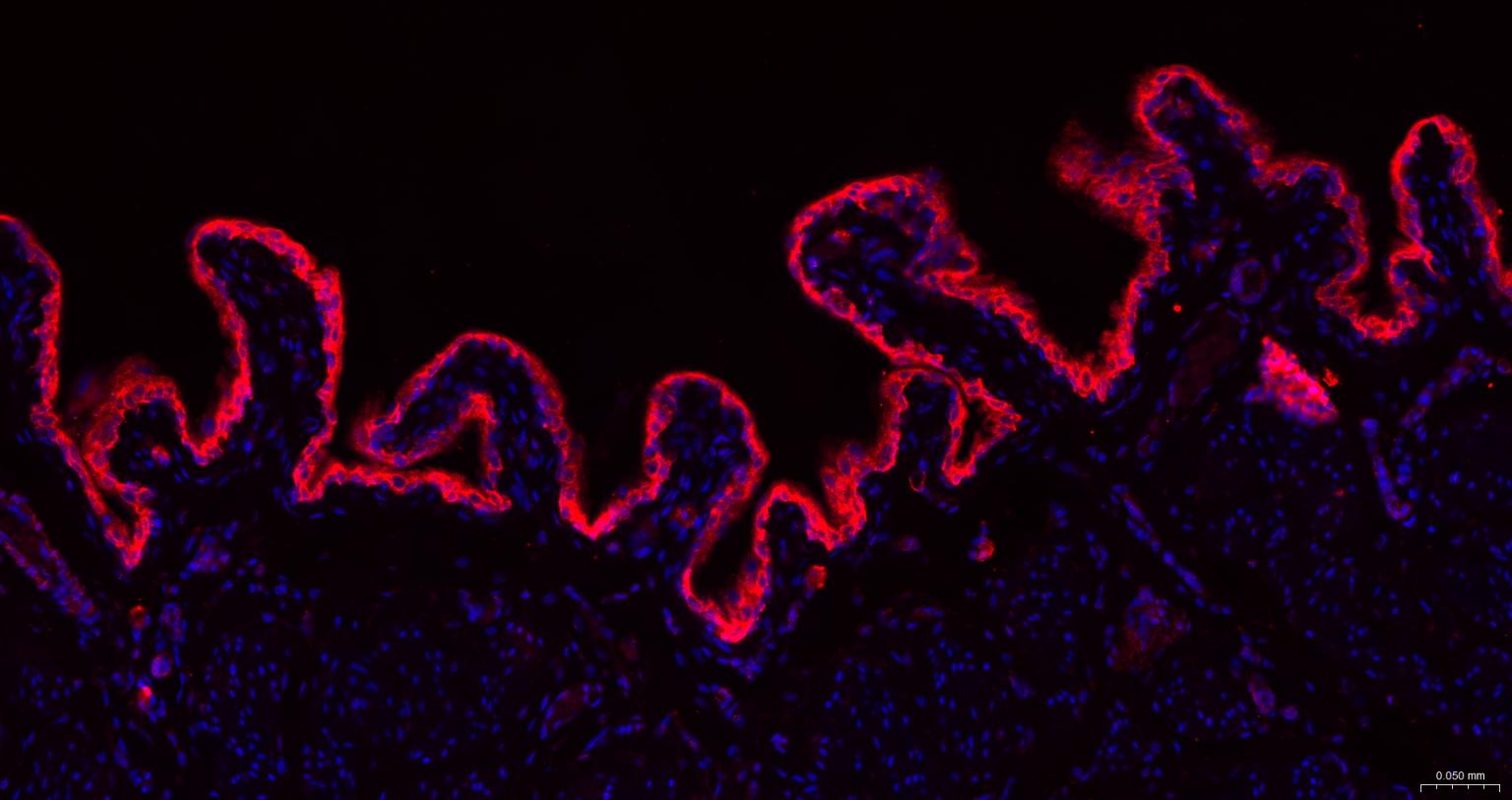
Paraformaldehyde-fixed, paraffin embedded Human Prostate; Antigen retrieval by boiling in sodium citrate buffer (pH6.0) for 15 min; The section was incubated with Pan Cytokeratin Polyclonal Antibody at 1:200 overnight at 4°C. Followed by conjugated Goat Anti-Rabbit IgG antibody (Red), DAPI (blue) was used to stain the cell nuclei.IF-P
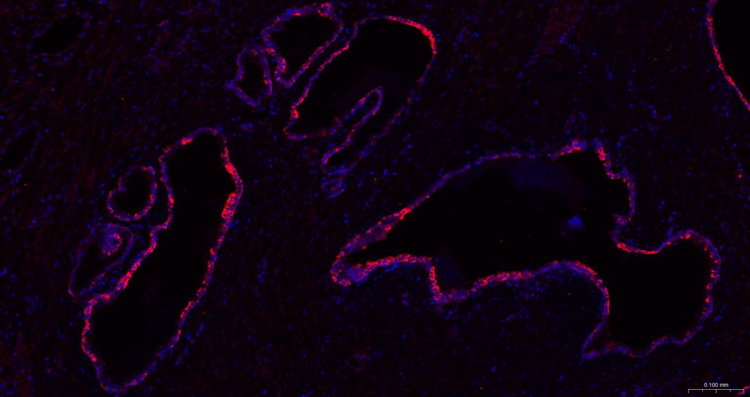
Paraformaldehyde-fixed, paraffin embedded Rat Bladder; Antigen retrieval by boiling in sodium citrate buffer (pH6.0) for 15 min; The section was incubated with Pan Cytokeratin Polyclonal Antibody at 1:200 overnight at 4°C. Followed by conjugated Goat Anti-Rabbit IgG antibody (Red), DAPI (blue) was used to stain the cell nuclei.FC
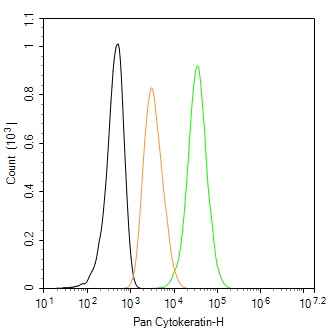
The HeLa (H) cells were fixed with 4% PFA (10 min at r.t.) and then permeabilized with 90% ice-cold methanol for 20 min at -20℃,the cells then were incubated in 5%BSA to block non-specific protein-protein interactions (30 min at r.t.). Cells stained with Primary Antibody for 30 min at room temperature,followed by secondary antibody incubation for 40 min at room temperature. Primary Antibody (green):Rabbit Anti-Pan Cytokeratin antibody (1:100); Isotype Control (orange): Rabbit IgG . Blank control (black): PBS. Acquisition of 20,000 events was performed.| Product Name | pan CK | pan Cytokeratin Rabbit pAb |
|---|---|
| Antibody Type | Primary Antibodies |
| Immunogen | KLH conjugated synthetic peptide derived from human Cytokeratin-1 : 141-240/644. |
| Clonality | Polyclonal |
|---|---|
| Isotype | IgG |
| Host Species | Rabbit |
| Tested Applications | FCICC/IFIF-PIHCWB |
| WB:1:500-1:2000 IHC:1:100-1:500 ICC/IF:1:100-1:500 IF-P:1:100-1:500 FC:1μg /test |
|
| Species Reactivity | HumanMouseRat |
| Concentration | 1mg/ml |
| Purification | Protein A |
| Gene Symbol | KRT13 |
|---|---|
| Gene Synonyms | K13 CK13 WSN2 |
| Gene Full Name | keratin 13 |
| Gene Summary | The protein encoded by this gene is a member of the keratin gene family. The keratins are intermediate filament proteins responsible for the structural integrity of epithelial cells and are subdivided into cytokeratins and hair keratins. Most of the type I cytokeratins consist of acidic proteins which are arranged in pairs of heterotypic keratin chains. This type I cytokeratin is paired with keratin 4 and expressed in the suprabasal layers of non-cornified stratified epithelia. Mutations in this gene and keratin 4 have been associated with the autosomal dominant disorder White Sponge Nevus. The type I cytokeratins are clustered in a region of chromosome 17q21.2. Alternative splicing of this gene results in multiple transcript variants; however, not all variants have been described. [provided by RefSeq, Jul 2008] |
| Alternative Names | CK13 K13 WSN2 K1C16_HUMAN KRT16 Cytokeratin-16 (CK-16) Keratin-16 (K16) KRT16A CK14 EBS1 EBS1A EBS1B EBS1C EBS1D EBS3 EBS4 K14 NFJ K1C14_HUMAN KRT14 Cytokeratin-14 (CK-14) Keratin-14 (K14) CK16 FNEPPK K16 K1CP NEPPK PC1 39.1 CK-17 K17 PC PC2 PCHC1 K1C17_HUMAN KRT17 Cytokeratin-17 (CK-17) Keratin-17 (K17) CK-24 K24 KA24 K1C24_HUMAN KRT24 Cytokeratin-24 (CK-24) Keratin-24 (K24) Type I keratin-24 keratin 24 keratin 24 type I pan-cytokeratin pan-CK pan CK P-CK CK-PAN wide spectrum Cytokeratin Cytokeratins |
| Molecular Weight(MW) | 46,60 kDa |
WB
Sample: Lane 1: Hela (Human) Cell Lysate at 30 ug, Lane 2: A549 (Human) Cell Lysate at 30 ug, Lane 3: A673 (Human) Cell Lysate at 30 ug, Primary: Anti-Pan Cytokeratin at 1/1000 dilution, Secondary: IRDye800CW Goat Anti-Rabbit IgG at 1/20000 dilution.
IHC
Paraformaldehyde-fixed, paraffin embedded (Human stomach carcinoma); Antigen retrieval by boiling in sodium citrate buffer (pH6.0) for 15min; Block endogenous peroxidase by 3% hydrogen peroxide for 20 minutes; Blocking buffer (normal goat serum) at 37°C for 30min; Antibody incubation with (Pan Cytokeratin) Polyclonal Antibody at 1:400 overnight at 4°C, followed by operating according to SP Kit(Rabbit) instructionsand DAB staining.
IHC
Paraformaldehyde-fixed, paraffin embedded Rat Uterus; Antigen retrieval by boiling in sodium citrate buffer (pH6.0) for 15 min; The section was incubated with Pan Cytokeratin Polyclonal Antibody, Unconjugated at 1:200 overnight at 4°C, followed by conjugation to the Goat anti-Rabbit IgG (H&L) pAb-HRP and DAB staining.
ICC/IF
Hela cell; 4% Paraformaldehyde-fixed; Triton X-100 at room temperature for 20 min; Blocking buffer (normal goat serum) at 37°C for 20 min; Antibody incubation with (Pan Cytokeratin) polyclonal Antibody 1:100, 90 minutes at 37°C; followed by a conjugated Goat Anti-Rabbit IgG antibody at 37°C for 90 minutes, DAPI (blue) was used to stain the cell nuclei.
IF-P
Paraformaldehyde-fixed, paraffin embedded Human Prostate; Antigen retrieval by boiling in sodium citrate buffer (pH6.0) for 15 min; The section was incubated with Pan Cytokeratin Polyclonal Antibody at 1:200 overnight at 4°C. Followed by conjugated Goat Anti-Rabbit IgG antibody (Red), DAPI (blue) was used to stain the cell nuclei.
IF-P
Paraformaldehyde-fixed, paraffin embedded Rat Bladder; Antigen retrieval by boiling in sodium citrate buffer (pH6.0) for 15 min; The section was incubated with Pan Cytokeratin Polyclonal Antibody at 1:200 overnight at 4°C. Followed by conjugated Goat Anti-Rabbit IgG antibody (Red), DAPI (blue) was used to stain the cell nuclei.
FC
The HeLa (H) cells were fixed with 4% PFA (10 min at r.t.) and then permeabilized with 90% ice-cold methanol for 20 min at -20℃,the cells then were incubated in 5%BSA to block non-specific protein-protein interactions (30 min at r.t.). Cells stained with Primary Antibody for 30 min at room temperature,followed by secondary antibody incubation for 40 min at room temperature. Primary Antibody (green):Rabbit Anti-Pan Cytokeratin antibody (1:100); Isotype Control (orange): Rabbit IgG . Blank control (black): PBS. Acquisition of 20,000 events was performed.| Application Notes | WB:1:500-1:2000 IHC:1:100-1:500 ICC/IF:1:100-1:500 IF-P:1:100-1:500 FC:1μg /test |
|---|
| Form | Liquid |
|---|---|
| Storage Instructions | Shipped at 4℃. Store at -20℃ for one year. Avoid repeated freeze/thaw cycles. |
| Storage Buffer | 0.01M TBS (pH7.4) with 1% BSA, 0.02% Proclin300 and 50% Glycerol. |


