WB
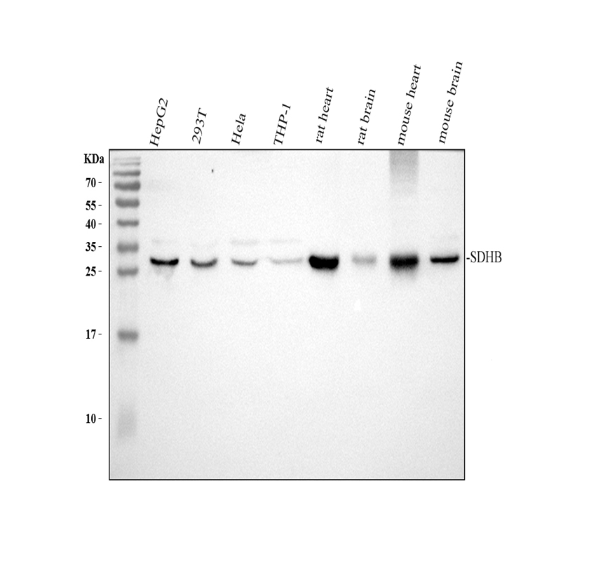
Western blot analysis of SDHB using anti-SDHB antibody. The sample well of each lane was loaded with 30 ug of sample under reducing conditions. After electrophoresis, proteins were transferred to a membrane. Then the membrane was incubated with rabbit anti-SDHB antigen affinity purified polyclonal antibody at a dilution of 1:1000 and probed with a goat anti-rabbit IgG-HRP secondary antibody. The signal is developed using ECL Plus Western Blotting Substrate.IHC
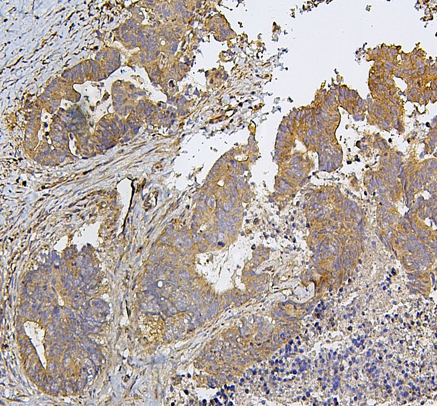
IHC analysis of SDHB using anti-SDHB antibody. SDHB was detected in a paraffin-embedded section of human colon cancer tissue. The tissue section was incubated with rabbit anti-SDHB Antibody at a dilution of 1:200 and developed using HRP Conjugated Rabbit IgG Super Vision Assay Kit with DAB as the chromogen.IHC
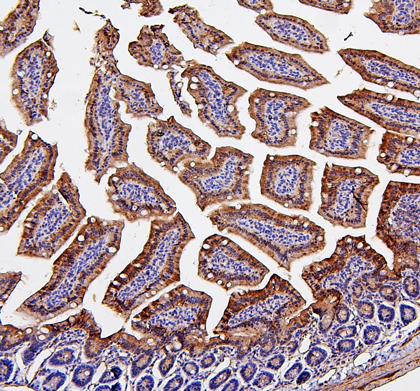
IHC analysis of SDHB using anti-SDHB antibody. SDHB was detected in a paraffin-embedded section of mouse small intestine tissue. The tissue section was incubated with rabbit anti-SDHB Antibody at a dilution of 1:200 and developed using HRP Conjugated Rabbit IgG Super Vision Assay Kit with DAB as the chromogen.IHC
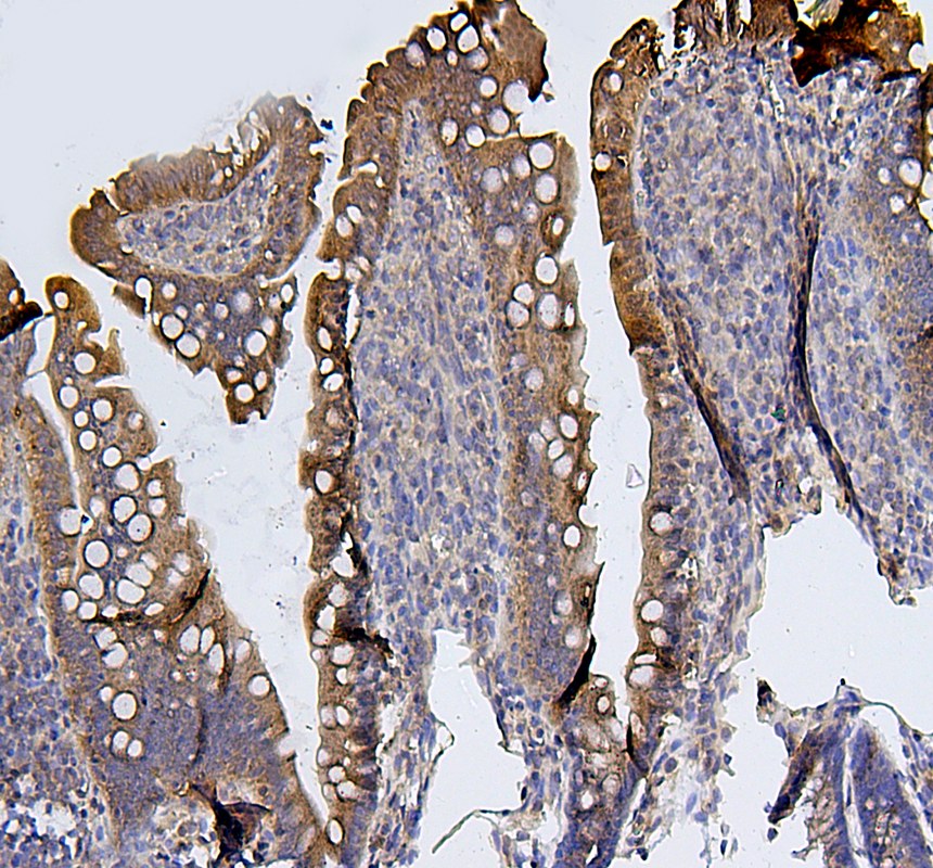
IHC analysis of SDHB using anti-SDHB antibody. SDHB was detected in a paraffin-embedded section of rat small intestine tissue. The tissue section was incubated with rabbit anti-SDHB Antibody at a dilution of 1:200 and developed using HRP Conjugated Rabbit IgG Super Vision Assay Kit with DAB as the chromogen.| Product Name | Rabbit polyclonal antibody to SDHB |
|---|---|
| Antibody Type | Primary Antibodies |
| Immunogen | E. coli-derived human SDHB recombinant protein (Position: A29-V280). Human SDHB shares 96.4% and 95.6% amino acid (aa) sequence identity with mouse and rat SDHB, respectively. |
| Clonality | Polyclonal |
|---|---|
| Isotype | IgG |
| Host Species | Rabbit |
| Tested Applications | IHCWB |
| WB:1:1000-1:8000 IHC:1:50-1:400 |
|
| Species Reactivity | HumanMouseRat |
| Concentration | 0.5mg/ml |
| Purification | Affinity purified |
| Gene Symbol | SDHB |
|---|---|
| Gene Synonyms | IP SDH CWS2 PGL4 SDH1 SDH2 PPGL4 SDHIP MC2DN4 |
| Gene Full Name | succinate dehydrogenase complex iron sulfur subunit B |
| Gene Summary | This tumor suppressor gene encodes the iron-sulfur protein subunit of the succinate dehydrogenase (SDH) enzyme complex which plays a critical role in mitochondria. The SDH enzyme complex is composed of four nuclear-encoded subunits. This enzyme complex converts succinate to fumarate which releases electrons as part of the citric acid cycle, and the enzyme complex additionally provides an attachment site for released electrons to be transferred to the oxidative phosphorylation pathway. The SDH enzyme complex plays a role in oxygen-related gene regulation through its conversion of succinate, which is an oxygen sensor that stabilizes the hypoxia-inducible factor 1 (HIF1) transcription factor. Sporadic and familial mutations in this gene result in paragangliomas, pheochromocytoma, and gastrointestinal stromal tumors, supporting a link between mitochondrial dysfunction and tumorigenesis. Mutations in this gene are also implicated in nuclear type 4 mitochondrial complex II deficiency. [provided by RefSeq, Jun 2022] |
| Alternative Names | IP PGL4 SDH SDH1 SDHB SDHIP |
| Molecular Weight(MW) | 32 kDa(Observed band size: 29 kDa) |
| Cellular Localization | Membrane,Mitochondrion,Mitochondrion inner membrane. |
WB
Western blot analysis of SDHB using anti-SDHB antibody. The sample well of each lane was loaded with 30 ug of sample under reducing conditions. After electrophoresis, proteins were transferred to a membrane. Then the membrane was incubated with rabbit anti-SDHB antigen affinity purified polyclonal antibody at a dilution of 1:1000 and probed with a goat anti-rabbit IgG-HRP secondary antibody. The signal is developed using ECL Plus Western Blotting Substrate.
IHC
IHC analysis of SDHB using anti-SDHB antibody. SDHB was detected in a paraffin-embedded section of human colon cancer tissue. The tissue section was incubated with rabbit anti-SDHB Antibody at a dilution of 1:200 and developed using HRP Conjugated Rabbit IgG Super Vision Assay Kit with DAB as the chromogen.
IHC
IHC analysis of SDHB using anti-SDHB antibody. SDHB was detected in a paraffin-embedded section of mouse small intestine tissue. The tissue section was incubated with rabbit anti-SDHB Antibody at a dilution of 1:200 and developed using HRP Conjugated Rabbit IgG Super Vision Assay Kit with DAB as the chromogen.
IHC
IHC analysis of SDHB using anti-SDHB antibody. SDHB was detected in a paraffin-embedded section of rat small intestine tissue. The tissue section was incubated with rabbit anti-SDHB Antibody at a dilution of 1:200 and developed using HRP Conjugated Rabbit IgG Super Vision Assay Kit with DAB as the chromogen.| Application Notes | WB:1:1000-1:8000 IHC:1:50-1:400 |
|---|
| Form | Liquid |
|---|---|
| Storage Instructions | 12 months from date of receipt,-20℃ as supplied. |
| Storage Buffer | 500 ug/ml antibody with PBS, 0.02% NaN3, 1 mg/ml BSA and 50% glycerol. |


