IHC
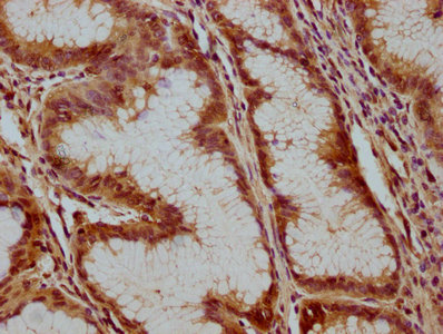
IHC image of ATP6V1C1 Antibody diluted at 1:100 and staining in paraffin-embedded human gastric cancer performed on a Leica BondTM system. After dewaxing and hydration, antigen retrieval was mediated by high pressure in a citrate buffer (pH 6.0). Section was blocked with 10% normal goat serum 30min at RT. Then primary antibody (1% BSA) was incubated at 4°C overnight. The primary is detected by a biotinylated secondary antibody and visualized using an HRP conjugated SP system.ICC/IF
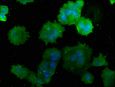
Immunofluorescence staining of MCF-7 cells with ATP6V1C1 Antibody at 1:50, counter-stained with DAPI. The cells were fixed in 4% formaldehyde, permeabilized using 0.2% Triton X-100 and blocked in 10% normal Goat Serum. The cells were then incubated with the antibody overnight at 4°C. The secondary antibody was Alexa Fluor 488-congugated AffiniPure Goat Anti-Rabbit IgG(H+L).| Product Name | Rabbit anti-ATP6V1C1 Polyclonal antibody |
|---|---|
| Antibody Type | Primary Antibodies |
| Immunogen | Recombinant Human V-type proton ATPase subunit C 1 protein (129-382AA). |
| Clonality | Polyclonal |
|---|---|
| Isotype | IgG |
| Host Species | Rabbit |
| Tested Applications | ICC/IFIHC |
| IHC:1:20-1:200 ICC/IF:1:50-1:200 |
|
| Species Reactivity | Human |
| Concentration | 1mg/ml |
| Purification | Affinity purified |
| Gene Symbol | ATP6V1C1 |
|---|---|
| Gene Synonyms | VATC Vma5 ATP6C ATP6D |
| Gene Full Name | ATPase H+ transporting V1 subunit C1 |
| Gene Summary | This gene encodes a component of vacuolar ATPase (V-ATPase), a multisubunit enzyme that mediates acidification of intracellular compartments of eukaryotic cells. V-ATPase dependent acidification is necessary for such intracellular processes as protein sorting, zymogen activation, receptor-mediated endocytosis, and synaptic vesicle proton gradient generation. V-ATPase is composed of a cytosolic V1 domain and a transmembrane V0 domain. The V1 domain consists of three A and three B subunits, two G subunits plus the C, D, E, F, and H subunits. The V1 domain contains the ATP catalytic site. The V0 domain consists of five different subunits: a, c, c', c'', and d. Additional isoforms of many of the V1 and V0 subunit proteins are encoded by multiple genes or alternatively spliced transcript variants. This gene is one of two genes that encode the V1 domain C subunit proteins and is found ubiquitously. This C subunit is analogous but not homologous to gamma subunit of F-ATPases. Previously, this gene was designated ATP6D. [provided by RefSeq, Jul 2008] |
| Alternative Names | ATP6C antibody ATP6D antibody ATP6V1C1 antibody ATPase H+ transporting lysosomal (vacuolar proton pump) 42kD antibody ATPase H+ transporting lysosomal 42kD V1 subunit C isoform 1 antibody ATPase H+ transporting lysosomal 42kDa V1 subunit C isoform 1 antibody ATPase H+ transporting lysosomal 42kDa V1 subunit C1 antibody ATPase H+ transporting lysosomal V1 subunit C1 antibody FLJ20057 antibody H(+) transporting two sector ATPase subunit C antibody H+ ATPase C subunit antibody H+ transporting ATPase chain C vacuolar antibody Subunit C of vacuolar proton ATPase V1 domain antibody V ATPase C subunit antibody V ATPase subunit C 1 antibody V-ATPase subunit C 1 antibody V-type proton ATPase subunit C 1 antibody Vacuolar ATP synthase subunit C antibody Vacuolar proton pump 42 kD subunit antibody Vacuolar proton pump C subunit antibody Vacuolar proton pump subunit C 1 antibody Vacuolar protonATPase subunit C VI domain antibody VATC antibody VATC1_HUMAN antibody VATPase C subunit antibody VATPase subunit C 1 antibody VMA5 antibody |
| Cellular Localization | Cytoplasmic vesicle,Membrane,Synapse. |
IHC
IHC image of ATP6V1C1 Antibody diluted at 1:100 and staining in paraffin-embedded human gastric cancer performed on a Leica BondTM system. After dewaxing and hydration, antigen retrieval was mediated by high pressure in a citrate buffer (pH 6.0). Section was blocked with 10% normal goat serum 30min at RT. Then primary antibody (1% BSA) was incubated at 4°C overnight. The primary is detected by a biotinylated secondary antibody and visualized using an HRP conjugated SP system.
ICC/IF
Immunofluorescence staining of MCF-7 cells with ATP6V1C1 Antibody at 1:50, counter-stained with DAPI. The cells were fixed in 4% formaldehyde, permeabilized using 0.2% Triton X-100 and blocked in 10% normal Goat Serum. The cells were then incubated with the antibody overnight at 4°C. The secondary antibody was Alexa Fluor 488-congugated AffiniPure Goat Anti-Rabbit IgG(H+L).| Application Notes | IHC:1:20-1:200 ICC/IF:1:50-1:200 |
|---|
| Form | Liquid |
|---|---|
| Storage Instructions | Upon receipt, store at -20°C or -80°C. Avoid repeated freeze. |
| Storage Buffer | Preservative: 0.03% Proclin 300; Constituents: 50% Glycerol, 0.01M PBS, pH 7.4. |


