WB
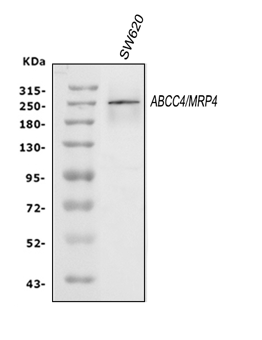
Western blot analysis of ABCC4 using anti-ABCC4 antibody. The sample well of each lane was loaded with 30 ug of sample under reducing conditions. After electrophoresis, proteins were transferred to a membrane. Then the membrane was incubated with rabbit anti-ABCC4 antigen affinity purified polyclonal antibody at a dilution of 1:1000 and probed with a goat anti-rabbit IgG-HRP secondary antibody. The signal is developed using ECL Plus Western Blotting Substrate.IHC
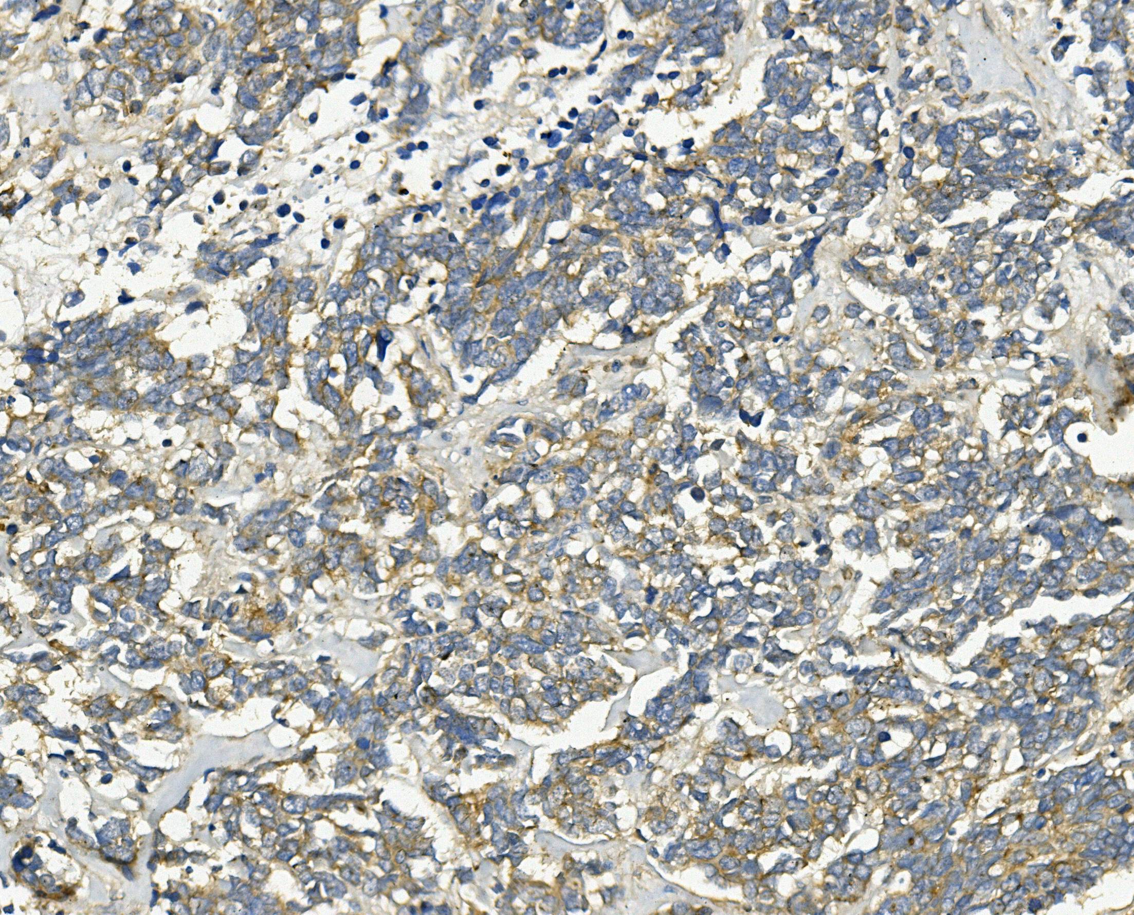
IHC analysis of ABCC4 using anti-ABCC4 antibody. ABCC4 was detected in a paraffin-embedded section of human lung cancer tissue. Biotinylated goat anti-rabbit IgG was used as secondary antibody. The tissue section was incubated with rabbit anti-ABCC4 Antibody at a dilution of 1:200 and developed using Strepavidin-Biotin-Complex (SABC) with DAB as the chromogen.ICC/IF
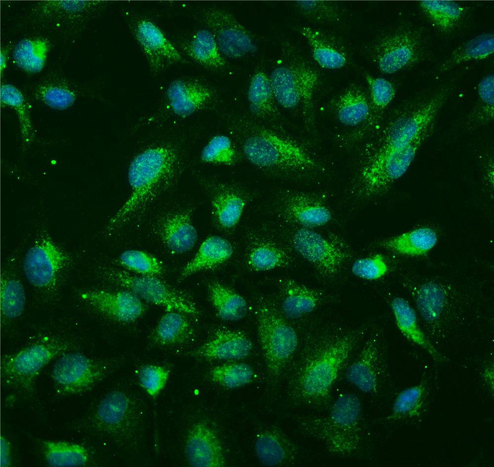
ICC/IF analysis of ABCC4 using anti-ABCC4-antibody. ABCC4 was detected in immunocytochemical section of A549 cells. Enzyme antigen retrieval was performed using IHC enzyme antigen retrieval reagent for 15 mins. The cells were blocked with 10% goat serum. And then incubated with 2μg/mL rabbit anti-ABCC4-Antibody overnight at 4°C. Fluoro488 Conjugated Goat Anti-Rabbit IgG was used as secondary antibody at 1:100 dilution and incubated for 30 minutes at 37°C. The section was counterstained with DAPI. Visualize using a fluorescence microscope and filter sets appropriate for the label used.FC
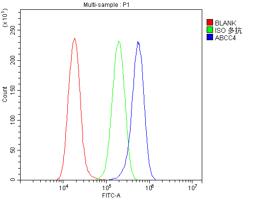
Flow Cytometry analysis of A549 cells using anti-ABCC4-antibody. Overlay histogram showing A549 cells stained with Anti-ABCC4 Antibody (Blue line). Anti-ABCC4-Antibody (1:100)) for 30 min at 20°C. Fluoro488 conjugated goat anti-rabbit IgG (1:100) was used as secondary antibody for 30 minutes at 20°C. Isotype control antibody (Green line) was rabbit IgG (1:100) used under the same conditions. Unlabelled sample (Red line) was also used as a control.| Product Name | Rabbit polyclonal antibody to ABCC4 |
|---|---|
| Antibody Type | Primary Antibodies |
| Immunogen | E.coli-derived human MRP4 recombinant protein (Position: M1-K77). |
| Clonality | Polyclonal |
|---|---|
| Isotype | IgG |
| Host Species | Rabbit |
| Tested Applications | FCICC/IFIHCWB |
| WB:1:500-1:2000 IHC:1:50-1:400 ICC/IF:1:50-1:400 FC:1:50-1:200 |
|
| Species Reactivity | Human |
| Concentration | 0.5mg/ml |
| Purification | Affinity purified |
| Gene Symbol | ABCC4 |
|---|---|
| Gene Synonyms | MRP4 MOATB MOAT-B |
| Gene Full Name | ATP binding cassette subfamily C member 4 (PEL blood group) |
| Gene Summary | The protein encoded by this gene is a member of the superfamily of ATP-binding cassette (ABC) transporters. ABC proteins transport various molecules across extra- and intra-cellular membranes. ABC genes are divided into seven distinct subfamilies (ABC1, MDR/TAP, MRP, ALD, OABP, GCN20, White). This protein is a member of the MRP subfamily which is involved in multi-drug resistance. This family member plays a role in cellular detoxification as a pump for its substrate, organic anions. It may also function in prostaglandin-mediated cAMP signaling in ciliogenesis. Alternative splicing of this gene results in multiple transcript variants. [provided by RefSeq, Sep 2014] |
| Alternative Names | ABCC4 EST170205 MOAT B MOATB MRP4 |
| Molecular Weight(MW) | 150 kDa(Observed band size: 200-250 kDa) |
| Cellular Localization | Cell membrane,Membrane. |
WB
Western blot analysis of ABCC4 using anti-ABCC4 antibody. The sample well of each lane was loaded with 30 ug of sample under reducing conditions. After electrophoresis, proteins were transferred to a membrane. Then the membrane was incubated with rabbit anti-ABCC4 antigen affinity purified polyclonal antibody at a dilution of 1:1000 and probed with a goat anti-rabbit IgG-HRP secondary antibody. The signal is developed using ECL Plus Western Blotting Substrate.
IHC
IHC analysis of ABCC4 using anti-ABCC4 antibody. ABCC4 was detected in a paraffin-embedded section of human lung cancer tissue. Biotinylated goat anti-rabbit IgG was used as secondary antibody. The tissue section was incubated with rabbit anti-ABCC4 Antibody at a dilution of 1:200 and developed using Strepavidin-Biotin-Complex (SABC) with DAB as the chromogen.
ICC/IF
ICC/IF analysis of ABCC4 using anti-ABCC4-antibody. ABCC4 was detected in immunocytochemical section of A549 cells. Enzyme antigen retrieval was performed using IHC enzyme antigen retrieval reagent for 15 mins. The cells were blocked with 10% goat serum. And then incubated with 2μg/mL rabbit anti-ABCC4-Antibody overnight at 4°C. Fluoro488 Conjugated Goat Anti-Rabbit IgG was used as secondary antibody at 1:100 dilution and incubated for 30 minutes at 37°C. The section was counterstained with DAPI. Visualize using a fluorescence microscope and filter sets appropriate for the label used.
FC
Flow Cytometry analysis of A549 cells using anti-ABCC4-antibody. Overlay histogram showing A549 cells stained with Anti-ABCC4 Antibody (Blue line). Anti-ABCC4-Antibody (1:100)) for 30 min at 20°C. Fluoro488 conjugated goat anti-rabbit IgG (1:100) was used as secondary antibody for 30 minutes at 20°C. Isotype control antibody (Green line) was rabbit IgG (1:100) used under the same conditions. Unlabelled sample (Red line) was also used as a control.| Application Notes | WB:1:500-1:2000 IHC:1:50-1:400 ICC/IF:1:50-1:400 FC:1:50-1:200 |
|---|
| Form | Liquid |
|---|---|
| Storage Instructions | 12 months from date of receipt,-20℃ as supplied. |
| Storage Buffer | 500 ug/ml antibody with PBS, 0.02% NaN3, 1 mg/ml BSA and 50% glycerol. |


