ICC/IF
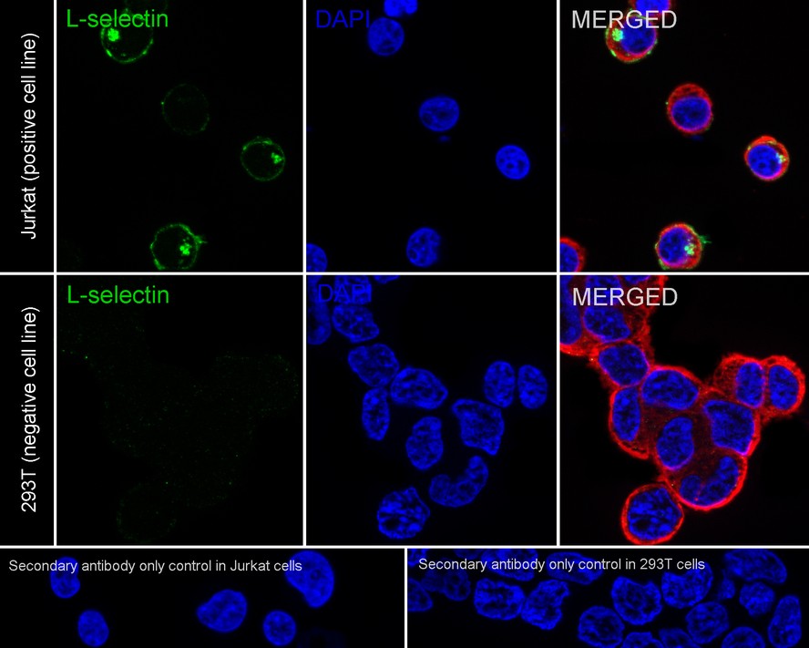
Immunocytochemistry analysis of Jurkat (positive) and 293T (negative) labeling CD62L with Rabbit anti-CD62L antibody at 1/50 dilution. Cells were fixed in 4% paraformaldehyde for 15 minutes at room temperature, permeabilized with 0.1% Triton X-100 in PBS for 15 minutes at room temperature, then blocked with 1% BSA in 10% negative goat serum for 1 hour at room temperature. Cells were then incubated with Rabbit anti-CD62L antibody at 1/50 dilution in 1% BSA in PBST overnight at 4 ℃. Goat Anti-Rabbit IgG H&L (488) was used as the secondary antibody at 1/1,000 dilution. PBS instead of the primary antibody was used as the secondary antibody only control. Nuclear DNA was labelled in blue with DAPI. Beta tubulin (red) was stained at 1/100 dilution overnight at +4℃. Goat Anti-Mouse IgG H&L (594) was used as the secondary antibody at 1/1,000 dilution.FC
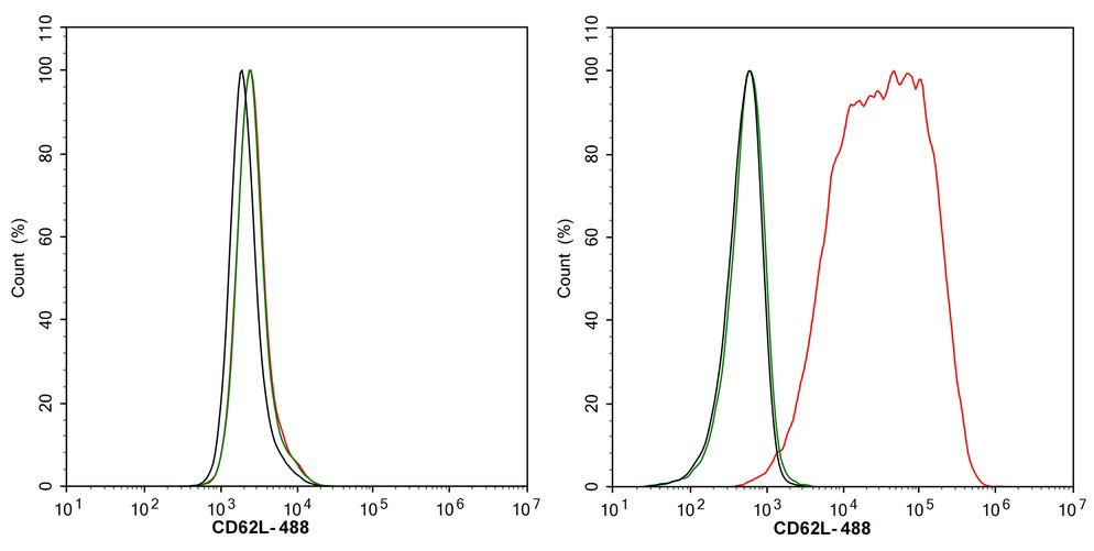
Flow cytometric analysis of 293T (left, negative) and Jurkat (right, positive) cells labeling CD62L. Cells were washed twice with cold PBS and resuspend. Then stained with the primary antibody (1/1,000) (red) compared with Rabbit IgG Isotype Control (green). After incubation of the primary antibody at +4℃ for an hour, the cells were stained with a 488 conjugate-Goat anti-Rabbit IgG Secondary antibody at 1/1,000 dilution for 30 minutes at +4℃. Unlabelled sample was used as a control (cells without incubation with primary antibody; black).| Product Name | CD62L Recombinant Rabbit Monoclonal Antibody |
|---|---|
| Antibody Type | Primary Antibodies |
| Immunogen | Recombinant protein within human CD62L aa 1-332. |
| Clonality | Monoclonal |
|---|---|
| Isotype | IgG |
| Host Species | Rabbit |
| Tested Applications | FCICC/IF |
| ICC/IF:1:50 FC:1:1000 |
|
| Species Reactivity | Human |
| Concentration | 1mg/ml |
| Purification | Protein A |
| Gene Symbol | SELL |
|---|---|
| Gene Synonyms | TQ1 LAM1 LEU8 LNHR LSEL CD62L LYAM1 PLNHR LECAM1 |
| Gene Full Name | selectin L |
| Gene Summary | This gene encodes a cell surface adhesion molecule that belongs to a family of adhesion/homing receptors. The encoded protein contains a C-type lectin-like domain, a calcium-binding epidermal growth factor-like domain, and two short complement-like repeats. The gene product is required for binding and subsequent rolling of leucocytes on endothelial cells, facilitating their migration into secondary lymphoid organs and inflammation sites. Single-nucleotide polymorphisms in this gene have been associated with various diseases including immunoglobulin A nephropathy. Alternatively spliced transcript variants have been found for this gene. [provided by RefSeq, Oct 2009] |
| Alternative Names | A.11 antibody AI528707 antibody CD62 antigen ligand antibody CD62 antigen-like family member L antibody CD62L antibody gp90-MEL antibody IgA nephropathy susceptibility to included antibody L Selectin antibody L-selectin antibody LAM-1 antibody LAM1 antibody LECAM1 antibody LEU8 antibody Leukocyte adhesion molecule 1 antibody Leukocyte surface antigen Leu-8 antibody Leukocyte-endothelial cell adhesion molecule 1 antibody Lnhr antibody LSEL antibody Ly-22 antibody Ly-m22 antibody Lyam-1 antibody LYAM1 antibody LYAM1_HUMAN antibody Lymph node homing receptor antibody Lymphocyte adhesion molecule 1 antibody Lymphocyte antigen 22 antibody Lymphocyte surface MEL-14 antigen antibody MEL-14 antibody Pln homing receptor antibody PLNHR antibody Selectin L antibody Selectin lymphocyte antibody SELL antibody TQ1 antibody |
| Molecular Weight(MW) | 42 kDa |
| Cellular Localization | Cell membrane. |
ICC/IF
Immunocytochemistry analysis of Jurkat (positive) and 293T (negative) labeling CD62L with Rabbit anti-CD62L antibody at 1/50 dilution. Cells were fixed in 4% paraformaldehyde for 15 minutes at room temperature, permeabilized with 0.1% Triton X-100 in PBS for 15 minutes at room temperature, then blocked with 1% BSA in 10% negative goat serum for 1 hour at room temperature. Cells were then incubated with Rabbit anti-CD62L antibody at 1/50 dilution in 1% BSA in PBST overnight at 4 ℃. Goat Anti-Rabbit IgG H&L (488) was used as the secondary antibody at 1/1,000 dilution. PBS instead of the primary antibody was used as the secondary antibody only control. Nuclear DNA was labelled in blue with DAPI. Beta tubulin (red) was stained at 1/100 dilution overnight at +4℃. Goat Anti-Mouse IgG H&L (594) was used as the secondary antibody at 1/1,000 dilution.
FC
Flow cytometric analysis of 293T (left, negative) and Jurkat (right, positive) cells labeling CD62L. Cells were washed twice with cold PBS and resuspend. Then stained with the primary antibody (1/1,000) (red) compared with Rabbit IgG Isotype Control (green). After incubation of the primary antibody at +4℃ for an hour, the cells were stained with a 488 conjugate-Goat anti-Rabbit IgG Secondary antibody at 1/1,000 dilution for 30 minutes at +4℃. Unlabelled sample was used as a control (cells without incubation with primary antibody; black).| Application Notes | ICC/IF:1:50 FC:1:1000 |
|---|
| Form | Liquid |
|---|---|
| Storage Instructions | 1*PBS (pH7.4). |
| Storage Buffer | Store at 2-8℃. Avoid freeze. |


