WB
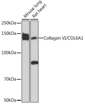
Western blot analysis of various lysates using Collagen VI/COL6A1 Rabbit mAb at 1:1000 dilution. Secondary antibody: HRP-conjugated Goat anti-Rabbit IgG (H+L) at 1:10000 dilution. Lysates/proteins: 25μg per lane. Blocking buffer: 3% nonfat dry milk in TBST. Detection: ECL Enhanced Kit. Exposure time: 3min.IHC
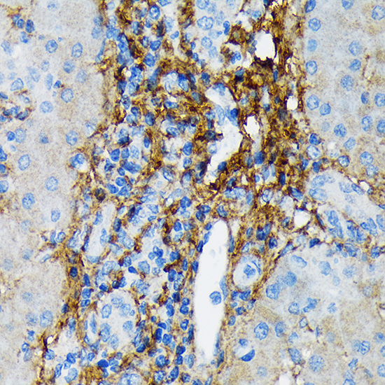
Immunohistochemistry analysis of paraffin embedded Rat lung using Collagen VI/COL6A1 Rabbit mAb at dilution of 1:100 (40x lens). Microwave antigen retrieval performed with 0.01M Tris/EDTA Buffer (pH 9.0) prior to IHC staining.IHC
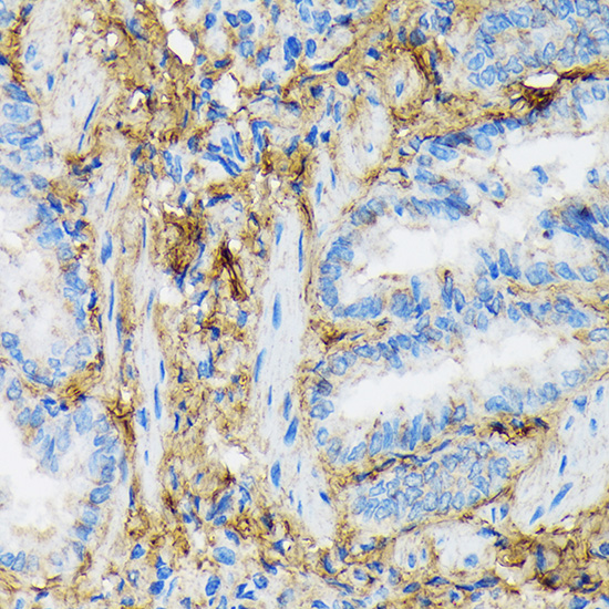
Immunohistochemistry analysis of paraffin embedded Human liver using Collagen VI/COL6A1 Rabbit mAb at dilution of 1:100 (40x lens). Microwave antigen retrieval performed with 0.01M Tris/EDTA Buffer (pH 9.0) prior to IHC staining.mIHC
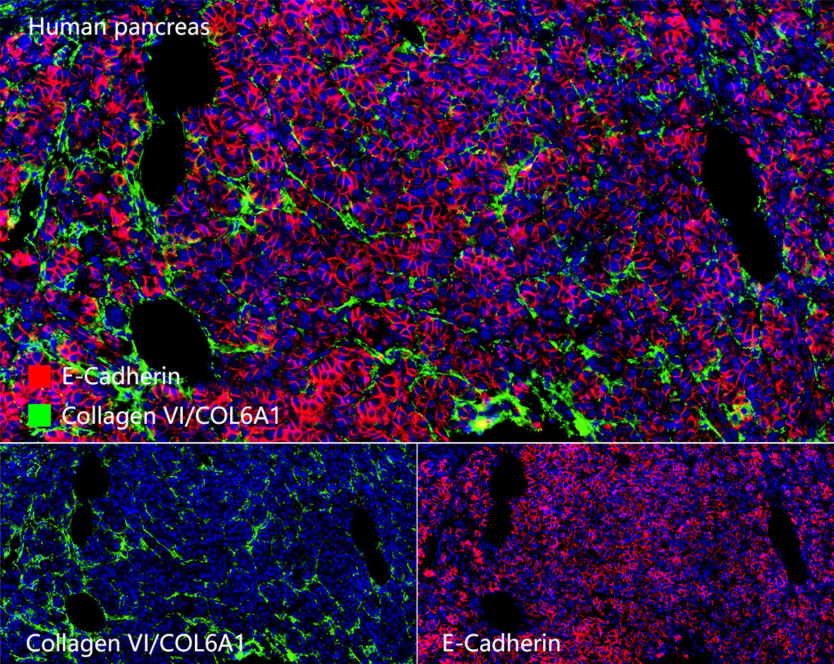
The multiplex IHC analysis on paraffin embedded Human tonsil tissue using the following specific primary antibodies and tyramide signal amplification (TSA) reagents : Collagen VI/COL6A1 Rabbit mAb (1:100) with TSA-TYR-520 (Green), and E-Cadherin Rabbit mAb (1:500) with TSA-TYR-570 (Red), and Cytokeratin 5 Rabbit mAb (1:2000) with TSA TYR-690 (Yellow). DAPI (Blue) was used for nuclear staining. Prior to multiplex IHC staining, high-pressure antigen retrieval was performed using 0.01M citrate buffer at pH 6.0. The analysis was completed using a 40x objective lens.| Product Name | Collagen VI/COL6A1 Rabbit mAb |
|---|---|
| Antibody Type | Primary Antibodies |
| Immunogen | Recombinant protein (or fragment).This information is considered to be commercially sensitive. |
| Clonality | Monoclonal |
|---|---|
| Isotype | IgG |
| Host Species | Rabbit |
| Tested Applications | IHCWBmIHC |
| WB:1:500-1:1000 IHC:1:50 - 1:200 mIHC:1:50 - 1:200 |
|
| Species Reactivity | HumanMouseRat |
| Concentration | 1mg/ml |
| Purification | Affinity purified |
| Gene Symbol | COL6A1 |
|---|---|
| Gene Synonyms | OPLL BTHLM1 UCHMD1 BTHLM1A UCHMD1A |
| Gene Full Name | collagen type VI alpha 1 chain |
| Gene Summary | The collagens are a superfamily of proteins that play a role in maintaining the integrity of various tissues. Collagens are extracellular matrix proteins and have a triple-helical domain as their common structural element. Collagen VI is a major structural component of microfibrils. The basic structural unit of collagen VI is a heterotrimer of the alpha1(VI), alpha2(VI), and alpha3(VI) chains. The alpha2(VI) and alpha3(VI) chains are encoded by the COL6A2 and COL6A3 genes, respectively. The protein encoded by this gene is the alpha 1 subunit of type VI collagen (alpha1(VI) chain). Mutations in the genes that code for the collagen VI subunits result in the autosomal dominant disorder, Bethlem myopathy. [provided by RefSeq, Jul 2008] |
| Alternative Names | OPLL BTHLM1 UCHMD1 Collagen VI/COL6A1 |
| Molecular Weight(MW) | 109 kDa(Observed band size: 140 kDa) |
| Cellular Localization | Secreted, extracellular matrix, extracellular space. |
WB
Western blot analysis of various lysates using Collagen VI/COL6A1 Rabbit mAb at 1:1000 dilution. Secondary antibody: HRP-conjugated Goat anti-Rabbit IgG (H+L) at 1:10000 dilution. Lysates/proteins: 25μg per lane. Blocking buffer: 3% nonfat dry milk in TBST. Detection: ECL Enhanced Kit. Exposure time: 3min.
IHC
Immunohistochemistry analysis of paraffin embedded Rat lung using Collagen VI/COL6A1 Rabbit mAb at dilution of 1:100 (40x lens). Microwave antigen retrieval performed with 0.01M Tris/EDTA Buffer (pH 9.0) prior to IHC staining.
IHC
Immunohistochemistry analysis of paraffin embedded Human liver using Collagen VI/COL6A1 Rabbit mAb at dilution of 1:100 (40x lens). Microwave antigen retrieval performed with 0.01M Tris/EDTA Buffer (pH 9.0) prior to IHC staining.
mIHC
The multiplex IHC analysis on paraffin embedded Human tonsil tissue using the following specific primary antibodies and tyramide signal amplification (TSA) reagents : Collagen VI/COL6A1 Rabbit mAb (1:100) with TSA-TYR-520 (Green), and E-Cadherin Rabbit mAb (1:500) with TSA-TYR-570 (Red), and Cytokeratin 5 Rabbit mAb (1:2000) with TSA TYR-690 (Yellow). DAPI (Blue) was used for nuclear staining. Prior to multiplex IHC staining, high-pressure antigen retrieval was performed using 0.01M citrate buffer at pH 6.0. The analysis was completed using a 40x objective lens.| Application Notes | WB:1:500-1:1000 IHC:1:50 - 1:200 mIHC:1:50 - 1:200 |
|---|
| Form | Liquid |
|---|---|
| Storage Instructions | Store at -20℃. |
| Storage Buffer | Buffer: PBS with 0.05% proclin300, 0.05% BSA, 50% glycerol, pH7.3. |


