WB
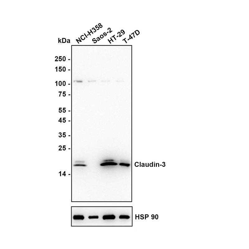
Western blot analysis of Claudin 3 on different lysates with Rabbit anti-Claudin 3 antibody at 1/50,000 dilution. Lysates/proteins at 10 µg/Lane. Exposure time: 30 seconds; Low expression of Claudin 3 protein in Saos-2 cell is consistent with the predicted expression pattern. Blocking: 5% NFDM/TBST, 1 hour at room temperature. Primary antibody: Claudin 3 Recombinant Rabbit Monoclonal Antibody, 1/50,000 in primary antibody dilution buffer, overnight at 4 °C. Secondary antibody: Goat anti-Rabbit IgG-HRP, 1/50,000 in 5% NFDM/TBST, 1 hour at room temperature.WB
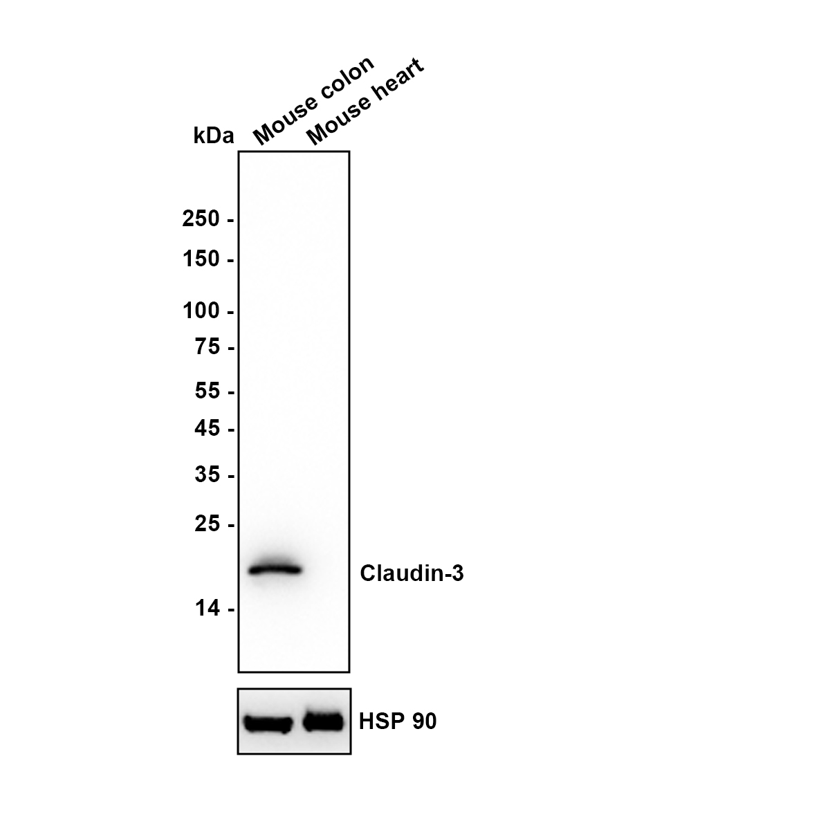
Western blot analysis of Claudin 3 on different lysates with Rabbit anti-Claudin 3 antibody at 1/50,000 dilution. Lysates/proteins at 20 µg/Lane. Exposure time: 3 seconds; Heart is a negative control. Blocking: 5% NFDM/TBST, 1 hour at room temperature. Primary antibody: Claudin 3 Recombinant Rabbit Monoclonal Antibody, 1/50,000 in primary antibody dilution buffer, overnight at 4 °C. Secondary antibody: Goat anti-Rabbit IgG-HRP, 1/50,000 in 5% NFDM/TBST, 1 hour at room temperature.IF-P
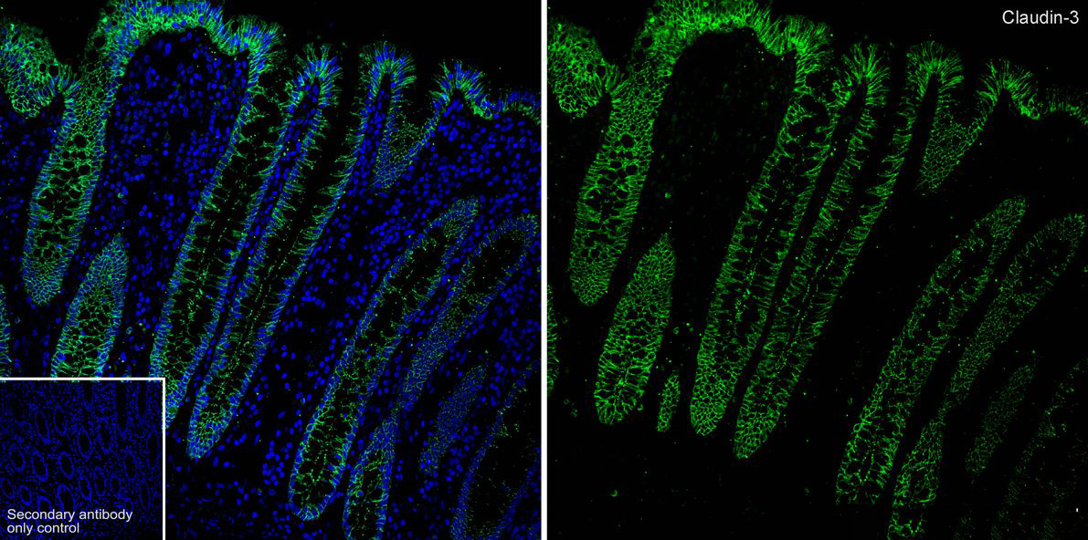
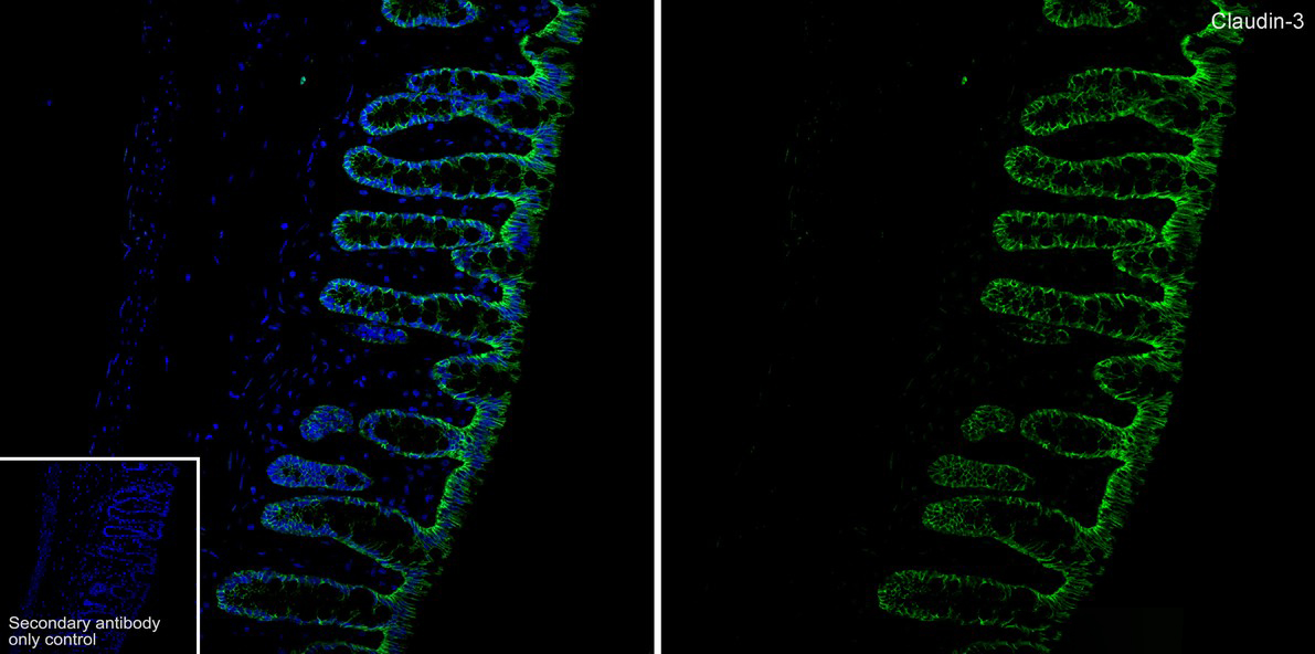
Application: Immunofluorescence (IF-tissue). Species: Human. Tissue: Colon. Sample: Paraffin-embedded section. Antigen retrieval: Heat-mediated, Tris-EDTA buffer (pH 9.0), 20 minutes at 95°C. Wash buffer: 1× TBST. Blocking: 10% normal goat serum + 1% Triton X-100 + 0.3 M Glycine in TBST, 30 minutes at room temperature. Primary antibody: Claudin 3 Recombinant Rabbit Monoclonal Antibody, 1/20,000, overnight at 4°C. Secondary antibody: Goat Anti-Rabbit IgG (488), 1.5 hours at room temperature.IF-P
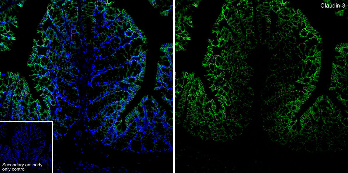
Application: Immunofluorescence (IF-tissue). Species: Mouse. Tissue: Colon. Sample: Paraffin-embedded section. Antigen retrieval: Heat-mediated, Tris-EDTA buffer (pH 9.0), 20 minutes at 95°C. Wash buffer: 1× TBST. Blocking: 10% normal goat serum + 1% Triton X-100 + 0.3 M Glycine in TBST, 30 minutes at room temperature. Primary antibody: Claudin 3 Recombinant Rabbit Monoclonal Antibody, 1/200, overnight at 4°C. Secondary antibody: Goat Anti-Rabbit IgG (488), 1.5 hours at room temperature.IF-P
Application: Immunofluorescence (IF-tissue). Species: Rat. Tissue: Colon. Sample: Paraffin-embedded section. Antigen retrieval: Heat-mediated, Tris-EDTA buffer (pH 9.0), 20 minutes at 95°C. Wash buffer: 1× TBST. Blocking: 10% normal goat serum + 1% Triton X-100 + 0.3 M Glycine in TBST, 30 minutes at room temperature. Primary antibody: Claudin 3 Recombinant Rabbit Monoclonal Antibody, 1/200, overnight at 4°C. Secondary antibody: Goat Anti-Rabbit IgG (488), 1.5 hours at room temperature.IP
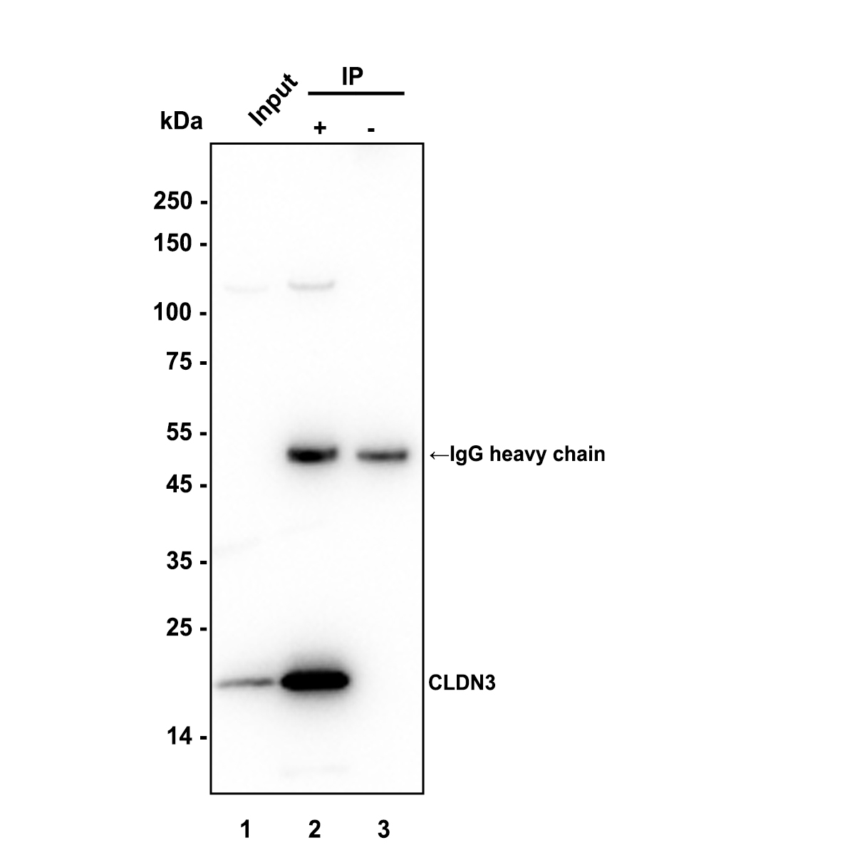
Immunoprecipitation (IP). Claudin 3 was immunoprecipitated in 0.2 mg MCF7 (Human breast cancer cells) cell lysate with Claudin 3 Recombinant Rabbit Monoclonal Antibodyat 2 µg/10 µl beads. Western blot was performed from the immunoprecipitate using Claudin 3 Recombinant Rabbit Monoclonal Antibodyat 1/20,000 dilution. Alpaca anti-Rabbit IgG Fc, Recombinant VHH monoclonal Antibody at 1/50,000 dilution was used for 1 hour at room temperature. Lane 1: MCF7 cell lysate (input). Lane 2: Claudin 3 Recombinant Rabbit Monoclonal AntibodyIP in MCF7 cell lysate. Lane 3: Rabbit IgG instead of Claudin 3 Recombinant Rabbit Monoclonal Antibodyin MCF7 cell lysate. Exposure time: 2 seconds. Blocking: 5% NFDM/TBST, 1 hour at room temperature. Primary dilution: Claudin 3 Recombinant Rabbit Monoclonal Antibody, 1/20,000 in primary antibody dilution buffer, 2 hours at room temperature.| Product Name | Claudin 3 Recombinant Rabbit Monoclonal Antibody |
|---|---|
| Antibody Type | Primary Antibodies |
| Immunogen | Synthetic peptide within human Claudin 3 aa 181-220. |
| Clonality | Monoclonal |
|---|---|
| Isotype | IgG |
| Host Species | Rabbit |
| Tested Applications | IF-PIPWB |
| WB:1:50000 IF-P:1:200-1:20000 IP:1-2μg/sample |
|
| Species Reactivity | HumanMouseRat |
| Concentration | 1mg/ml |
| Purification | Protein A |
| Gene Symbol | CLDN3 |
|---|---|
| Gene Synonyms | RVP1 HRVP1 C7orf1 CPE-R2 CPETR2 |
| Gene Full Name | claudin 3 |
| Gene Summary | Tight junctions represent one mode of cell-to-cell adhesion in epithelial or endothelial cell sheets, forming continuous seals around cells and serving as a physical barrier to prevent solutes and water from passing freely through the paracellular space. These junctions are comprised of sets of continuous networking strands in the outwardly facing cytoplasmic leaflet, with complementary grooves in the inwardly facing extracytoplasmic leaflet. The protein encoded by this intronless gene, a member of the claudin family, is an integral membrane protein and a component of tight junction strands. It is also a low-affinity receptor for Clostridium perfringens enterotoxin, and shares aa sequence similarity with a putative apoptosis-related protein found in rat. [provided by RefSeq, Jul 2008] |
| Alternative Names | C7orf1 antibody Claudin-3 antibody Claudin3 antibody CLD3_HUMAN antibody CLDN 3 antibody Cldn3 antibody Clostridium perfringens enterotoxin receptor 2 antibody CPE R2 antibody CPE receptor 2 antibody CPE-R 2 antibody CPE-receptor 2 antibody CPETR 2 antibody CPETR2 antibody HRVP 1 antibody HRVP1 antibody Rat ventral prostate 1 like protein antibody Rat ventral prostate.1 protein homolog antibody RVP1 antibody Ventral prostate.1 like protein antibody Ventral prostate.1 protein homolog antibody |
| Molecular Weight(MW) | 23 kDa(Observed band size: 20 kDa) |
| Cellular Localization | Cell junction, tight junction, Cell membrane. |
WB
Western blot analysis of Claudin 3 on different lysates with Rabbit anti-Claudin 3 antibody at 1/50,000 dilution. Lysates/proteins at 10 µg/Lane. Exposure time: 30 seconds; Low expression of Claudin 3 protein in Saos-2 cell is consistent with the predicted expression pattern. Blocking: 5% NFDM/TBST, 1 hour at room temperature. Primary antibody: Claudin 3 Recombinant Rabbit Monoclonal Antibody, 1/50,000 in primary antibody dilution buffer, overnight at 4 °C. Secondary antibody: Goat anti-Rabbit IgG-HRP, 1/50,000 in 5% NFDM/TBST, 1 hour at room temperature.
WB
Western blot analysis of Claudin 3 on different lysates with Rabbit anti-Claudin 3 antibody at 1/50,000 dilution. Lysates/proteins at 20 µg/Lane. Exposure time: 3 seconds; Heart is a negative control. Blocking: 5% NFDM/TBST, 1 hour at room temperature. Primary antibody: Claudin 3 Recombinant Rabbit Monoclonal Antibody, 1/50,000 in primary antibody dilution buffer, overnight at 4 °C. Secondary antibody: Goat anti-Rabbit IgG-HRP, 1/50,000 in 5% NFDM/TBST, 1 hour at room temperature.
IF-P
Application: Immunofluorescence (IF-tissue). Species: Human. Tissue: Colon. Sample: Paraffin-embedded section. Antigen retrieval: Heat-mediated, Tris-EDTA buffer (pH 9.0), 20 minutes at 95°C. Wash buffer: 1× TBST. Blocking: 10% normal goat serum + 1% Triton X-100 + 0.3 M Glycine in TBST, 30 minutes at room temperature. Primary antibody: Claudin 3 Recombinant Rabbit Monoclonal Antibody, 1/20,000, overnight at 4°C. Secondary antibody: Goat Anti-Rabbit IgG (488), 1.5 hours at room temperature.
IF-P
Application: Immunofluorescence (IF-tissue). Species: Mouse. Tissue: Colon. Sample: Paraffin-embedded section. Antigen retrieval: Heat-mediated, Tris-EDTA buffer (pH 9.0), 20 minutes at 95°C. Wash buffer: 1× TBST. Blocking: 10% normal goat serum + 1% Triton X-100 + 0.3 M Glycine in TBST, 30 minutes at room temperature. Primary antibody: Claudin 3 Recombinant Rabbit Monoclonal Antibody, 1/200, overnight at 4°C. Secondary antibody: Goat Anti-Rabbit IgG (488), 1.5 hours at room temperature.
IF-P
Application: Immunofluorescence (IF-tissue). Species: Rat. Tissue: Colon. Sample: Paraffin-embedded section. Antigen retrieval: Heat-mediated, Tris-EDTA buffer (pH 9.0), 20 minutes at 95°C. Wash buffer: 1× TBST. Blocking: 10% normal goat serum + 1% Triton X-100 + 0.3 M Glycine in TBST, 30 minutes at room temperature. Primary antibody: Claudin 3 Recombinant Rabbit Monoclonal Antibody, 1/200, overnight at 4°C. Secondary antibody: Goat Anti-Rabbit IgG (488), 1.5 hours at room temperature.
IP
Immunoprecipitation (IP). Claudin 3 was immunoprecipitated in 0.2 mg MCF7 (Human breast cancer cells) cell lysate with Claudin 3 Recombinant Rabbit Monoclonal Antibodyat 2 µg/10 µl beads. Western blot was performed from the immunoprecipitate using Claudin 3 Recombinant Rabbit Monoclonal Antibodyat 1/20,000 dilution. Alpaca anti-Rabbit IgG Fc, Recombinant VHH monoclonal Antibody at 1/50,000 dilution was used for 1 hour at room temperature. Lane 1: MCF7 cell lysate (input). Lane 2: Claudin 3 Recombinant Rabbit Monoclonal AntibodyIP in MCF7 cell lysate. Lane 3: Rabbit IgG instead of Claudin 3 Recombinant Rabbit Monoclonal Antibodyin MCF7 cell lysate. Exposure time: 2 seconds. Blocking: 5% NFDM/TBST, 1 hour at room temperature. Primary dilution: Claudin 3 Recombinant Rabbit Monoclonal Antibody, 1/20,000 in primary antibody dilution buffer, 2 hours at room temperature.| Application Notes | WB:1:50000 IF-P:1:200-1:20000 IP:1-2μg/sample |
|---|
| Form | Liquid |
|---|---|
| Storage Instructions | Shipped at 4℃. Store at +4℃ short term (1-2 weeks). Store at -20℃ long term. |
| Storage Buffer | 1*PBS (pH7.4), 0.1% BSA, 40% Glycerol, 0.2% Proclean 950. |


