WB
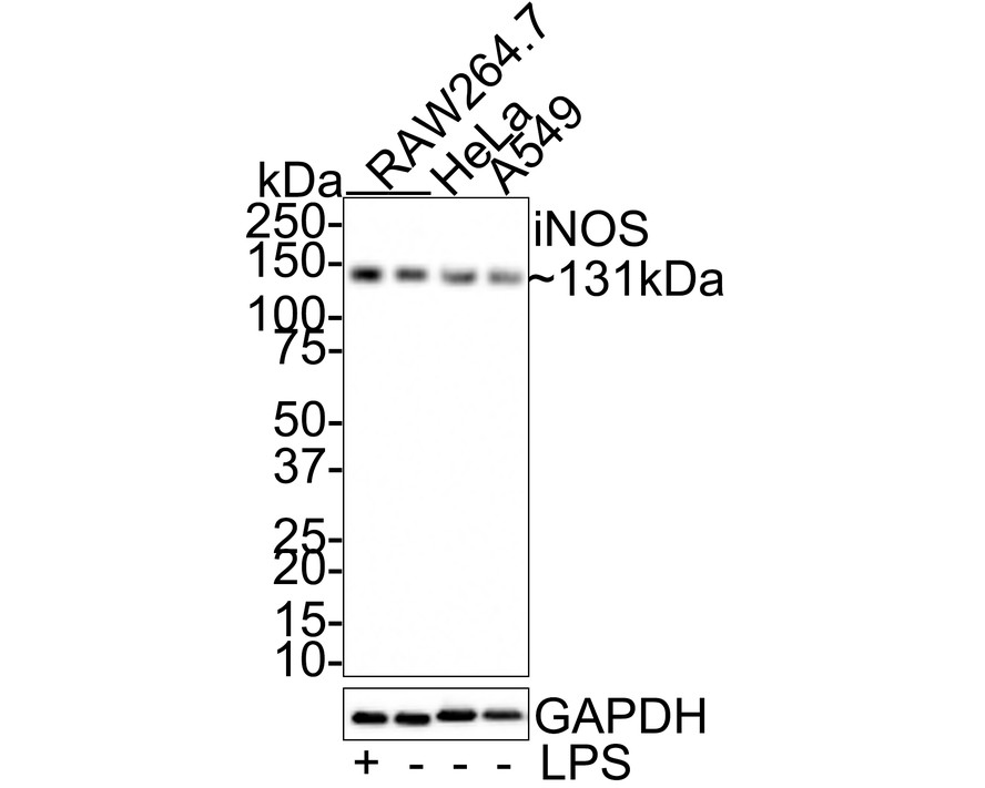
Western blot analysis of iNOS on different lysates with Rabbit anti-iNOS antibody at 1/1,000 dilution. Exposure time: 16 seconds; 4-20% SDS-PAGE gel. Proteins were transferred to a PVDF membrane and blocked with 5% NFDM/TBST for 1 hour at room temperature. The primary antibody at 1/1,000 dilution was used in 5% NFDM/TBST at room temperature for 2 hours. Goat Anti-Rabbit IgG - HRP Secondary Antibody at 1:100,000 dilution was used for 1 hour at room temperature.ICC/IF
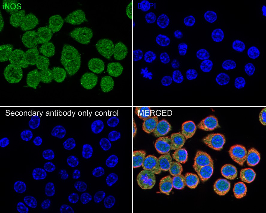
Immunocytochemistry analysis of RAW264.7 cells labeling iNOS with Rabbit anti-iNOS antibody at 1/100 dilution. Cells were fixed in 4% paraformaldehyde for 20 minutes at room temperature, permeabilized with 0.1% Triton X-100 in PBS for 5 minutes at room temperature, then blocked with 1% BSA in 10% negative goat serum for 1 hour at room temperature. Cells were then incubated with Rabbit anti-iNOS antibody at 1/100 dilution in 1% BSA in PBST overnight at 4 ℃. Goat Anti-Rabbit IgG H&L (488) was used as the secondary antibody at 1/1,000 dilution. PBS instead of the primary antibody was used as the secondary antibody only control. Nuclear DNA was labelled in blue with DAPI. Beta tubulin (red) was stained at 1/100 dilution overnight at +4℃. Goat Anti-Mouse IgG H&L (594) was used as the secondary antibody at 1/1,000 dilution.IF-F
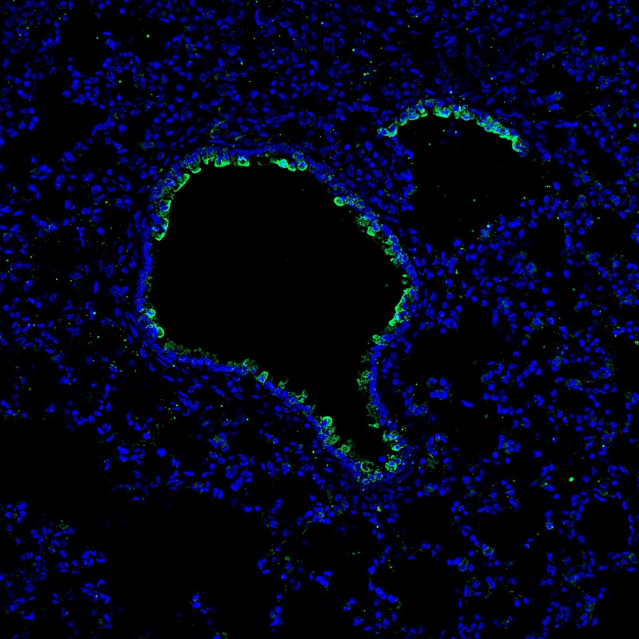
Application: IHC-Fr. Species: Mouse. Site: lung. Sample: Frozen section. Antibody concentration: 1/100. Antigen retrieval: Not required.| Product Name | iNOS Rabbit Polyclonal Antibody |
|---|---|
| Antibody Type | Primary Antibodies |
| Immunogen | Synthetic peptide within human iNOS aa 1,104-1,153 / 1,153. |
| Clonality | Polyclonal |
|---|---|
| Isotype | IgG |
| Host Species | Rabbit |
| Tested Applications | ICC/IFIF-FWB |
| WB:1:2000-1:5000 ICC/IF:1:50-1:200 IF-F:1:100 |
|
| Species Reactivity | HumanMouseRat |
| Concentration | 1mg/ml |
| Purification | Affinity purified |
| Gene Symbol | NOS2 |
|---|---|
| Gene Synonyms | NOS INOS NOS2A HEP-NOS |
| Gene Full Name | nitric oxide synthase 2 |
| Gene Summary | Nitric oxide is a reactive free radical which acts as a biologic mediator in several processes, including neurotransmission and antimicrobial and antitumoral activities. This gene encodes a nitric oxide synthase which is expressed in liver and is inducible by a combination of lipopolysaccharide and certain cytokines. Three related pseudogenes are located within the Smith-Magenis syndrome region on chromosome 17. [provided by RefSeq, Jul 2008] |
| Alternative Names | HEP-NOS antibody Hepatocyte NOS antibody HEPNOS antibody inducible antibody Inducible nitric oxide synthase antibody Inducible NO synthase antibody Inducible NOS antibody iNOS antibody MAC NOS antibody Macrophage NOS antibody Nitric oxide synthase 2 inducible antibody Nitric oxide synthase 2 inducible macrophage antibody nitric oxide synthase 2A (inducible hepatocytes) antibody Nitric oxide synthase antibody Nitric oxide synthase inducible antibody nitric oxide synthase macrophage antibody NOS 2 antibody NOS antibody Nos II antibody NOS type II antibody nos2 antibody NOS2_HUMAN antibody NOS2A antibody NOS2A Inducible Hepatocyte antibody Peptidyl-cysteine S-nitrosylase NOS2 antibody |
| Molecular Weight(MW) | 131 kDa |
| Cellular Localization | Cytoskeleton, Nucleus, Cytosol. |
| SwissProt ID | P35228 |
|---|
WB
Western blot analysis of iNOS on different lysates with Rabbit anti-iNOS antibody at 1/1,000 dilution. Exposure time: 16 seconds; 4-20% SDS-PAGE gel. Proteins were transferred to a PVDF membrane and blocked with 5% NFDM/TBST for 1 hour at room temperature. The primary antibody at 1/1,000 dilution was used in 5% NFDM/TBST at room temperature for 2 hours. Goat Anti-Rabbit IgG - HRP Secondary Antibody at 1:100,000 dilution was used for 1 hour at room temperature.
ICC/IF
Immunocytochemistry analysis of RAW264.7 cells labeling iNOS with Rabbit anti-iNOS antibody at 1/100 dilution. Cells were fixed in 4% paraformaldehyde for 20 minutes at room temperature, permeabilized with 0.1% Triton X-100 in PBS for 5 minutes at room temperature, then blocked with 1% BSA in 10% negative goat serum for 1 hour at room temperature. Cells were then incubated with Rabbit anti-iNOS antibody at 1/100 dilution in 1% BSA in PBST overnight at 4 ℃. Goat Anti-Rabbit IgG H&L (488) was used as the secondary antibody at 1/1,000 dilution. PBS instead of the primary antibody was used as the secondary antibody only control. Nuclear DNA was labelled in blue with DAPI. Beta tubulin (red) was stained at 1/100 dilution overnight at +4℃. Goat Anti-Mouse IgG H&L (594) was used as the secondary antibody at 1/1,000 dilution.
IF-F
Application: IHC-Fr. Species: Mouse. Site: lung. Sample: Frozen section. Antibody concentration: 1/100. Antigen retrieval: Not required.| Application Notes | WB:1:2000-1:5000 ICC/IF:1:50-1:200 IF-F:1:100 |
|---|
| Form | Liquid |
|---|---|
| Storage Instructions | Shipped at 4℃. Store at +4℃ short term (1-2 weeks). It is recommended to aliquot into single-use upon delivery. Store at -20℃ long term. |
| Storage Buffer | 1*PBS (pH7.4), 0.2% BSA, 50% Glycerol. Preservative: 0.05% Sodium Azide. |


