WB
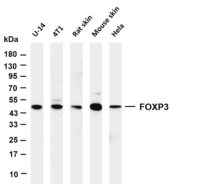
Various whole cell lysates were separated by 4-20% SDS-PAGE, and the membrane was blotted with anti-FOXP3 antibody. The HRP-conjugated Goat anti-Rabbit IgG (H + L) antibody was used to detect the antibody.IHC
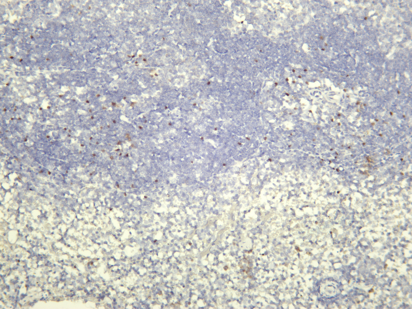
Mouse spleen was stained with Anti-FOXP3 rabbit antibody.IHC
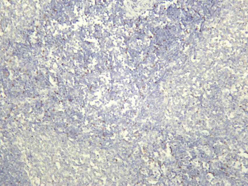
Rat spleen was stained with Anti-FOXP3 rabbit antibody.IHC
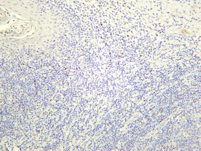
Human tonsil was stained with Anti-FOXP3 rabbit antibody.IF-P
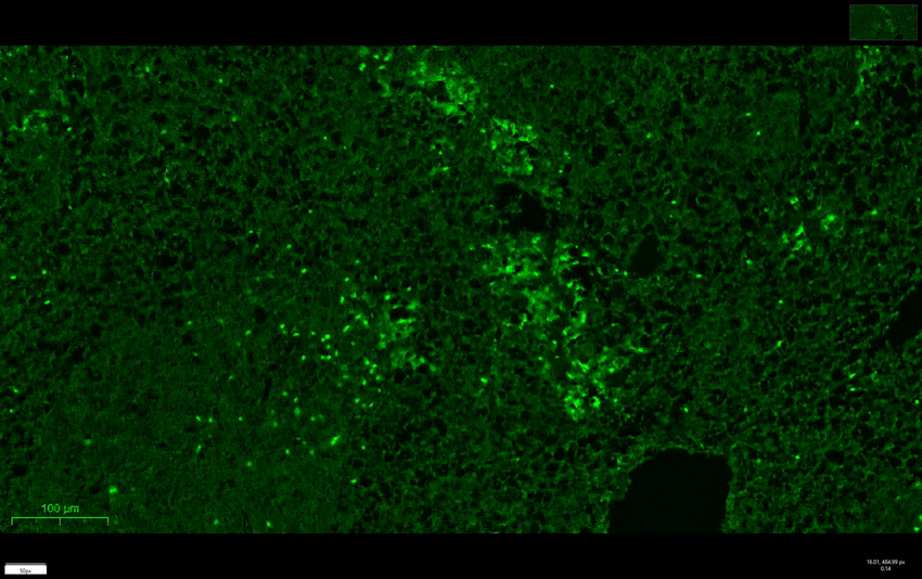
mouse spleen was stained with Anti-FOXP3 rabbit antibody.IF-P
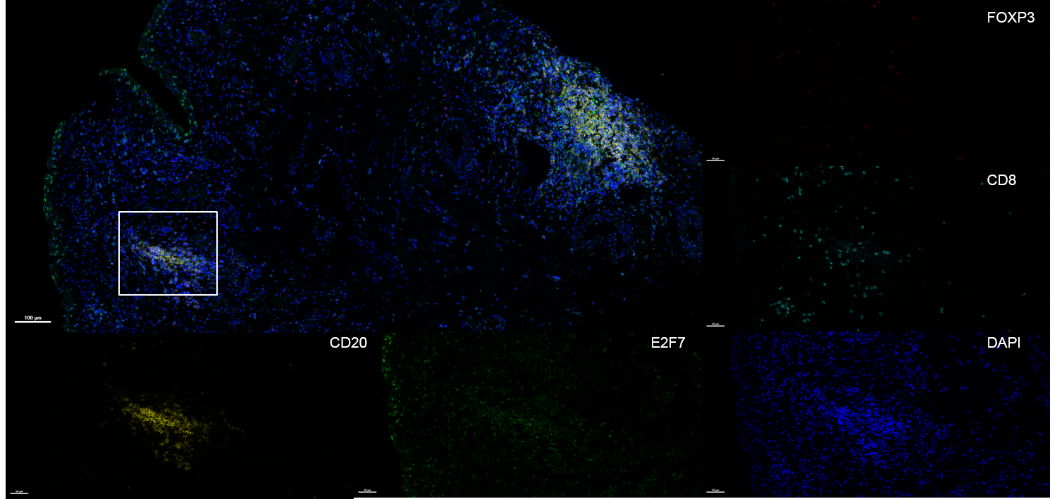
Fluorescence multiplex immunohistochemical analysis of Human bladder tissue (formalin-fixed paraffin-embedded section). The immunostaining was performed by Sextuple-Fluorescence kit. CD8 rabbit mAb cyan, FOXP3 rabbit mAb red,Neurofilament mouse mAb purple,Iba 1 mouse mAb red, CD20 mouse mAb yellow, The section was incubated in 4 rounds of staining; sequentially for Anti-antibodies; each using a separate fluorescent tyramide signal amplification system. EDTA based antigen retrieval (pH 9.0, 20 minutes) was used in between rounds of tyramide signal amplification to remove the antibody from the previous round, to avoid any cross-reactivity. DAPI (dark blue) was used as a nuclear counter stain. Microscopy and pseudocoloring of individual dyes was performed using a Slideviewer Imaging System (Excilone).| Product Name | FOXP3 Rabbit mAb |
|---|---|
| Antibody Type | Primary Antibodies |
| Immunogen | The specific immunogen used to produce this antibody is proprietary information. |
| Clonality | Monoclonal |
|---|---|
| Isotype | IgG,K |
| Host Species | Rabbit |
| Tested Applications | IF-PIHCWB |
| WB:1:1000-1:5000 IHC:1:200-1:1000 IF-P:1:200-1:1000 |
|
| Species Reactivity | HumanMousePigRat |
| Concentration | 1mg/ml |
| Purification | Protein A |
| Gene Symbol | FOXP3 |
|---|---|
| Gene Synonyms | JM2 AIID IPEX PIDX XPID DIETER |
| Gene Full Name | forkhead box P3 |
| Gene Summary | The protein encoded by this gene is a member of the forkhead/winged-helix family of transcriptional regulators. Defects in this gene are the cause of immunodeficiency polyendocrinopathy, enteropathy, X-linked syndrome (IPEX), also known as X-linked autoimmunity-immunodeficiency syndrome. Alternatively spliced transcript variants encoding different isoforms have been identified. [provided by RefSeq, Jul 2008] |
| Alternative Names | FOXP3 IPEX JM2 Forkhead box protein P3 Scurfin |
| Molecular Weight(MW) | 47 kDa |
| Cellular Localization | Nucleus. |
| SwissProt ID | Q9BZS1 |
|---|
WB
Various whole cell lysates were separated by 4-20% SDS-PAGE, and the membrane was blotted with anti-FOXP3 antibody. The HRP-conjugated Goat anti-Rabbit IgG (H + L) antibody was used to detect the antibody.
IHC
Mouse spleen was stained with Anti-FOXP3 rabbit antibody.
IHC
Rat spleen was stained with Anti-FOXP3 rabbit antibody.
IHC
Human tonsil was stained with Anti-FOXP3 rabbit antibody.
IF-P
mouse spleen was stained with Anti-FOXP3 rabbit antibody.
IF-P
Fluorescence multiplex immunohistochemical analysis of Human bladder tissue (formalin-fixed paraffin-embedded section). The immunostaining was performed by Sextuple-Fluorescence kit. CD8 rabbit mAb cyan, FOXP3 rabbit mAb red,Neurofilament mouse mAb purple,Iba 1 mouse mAb red, CD20 mouse mAb yellow, The section was incubated in 4 rounds of staining; sequentially for Anti-antibodies; each using a separate fluorescent tyramide signal amplification system. EDTA based antigen retrieval (pH 9.0, 20 minutes) was used in between rounds of tyramide signal amplification to remove the antibody from the previous round, to avoid any cross-reactivity. DAPI (dark blue) was used as a nuclear counter stain. Microscopy and pseudocoloring of individual dyes was performed using a Slideviewer Imaging System (Excilone).| Application Notes | WB:1:1000-1:5000 IHC:1:200-1:1000 IF-P:1:200-1:1000 |
|---|
| Form | Liquid |
|---|---|
| Storage Instructions | -15°C to -25°C/1 year (Do not lower than -25°C). |
| Storage Buffer | PBS, 50% glycerol, 0.05% Proclin 300, 0.05%BSA. |


