WB
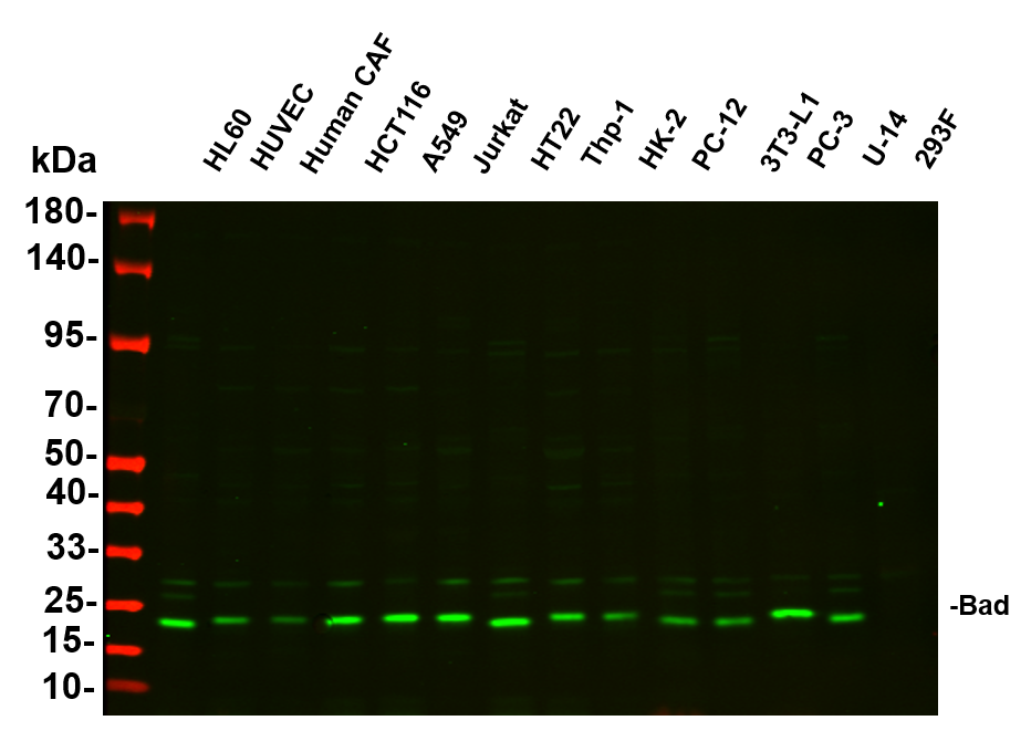
Various whole cell lysates were separated by 4-20% SDS-PAGE, and the primary antibody was used at 4℃ over night with a 1:2500 dilution . The Dilight 800-conjugated Goat anti-Rabbit antibody was used to detect the antibody.WB
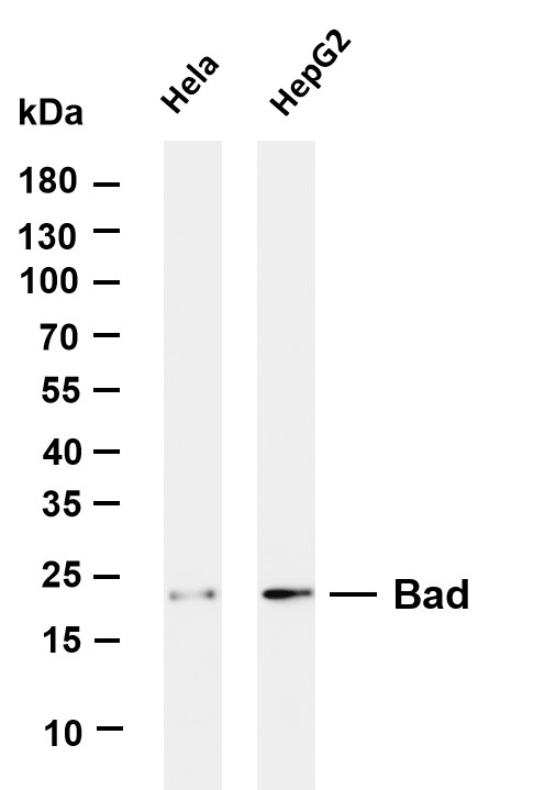
Various whole cell lysates were separated by 4-20% SDS-PAGE, and the membrane was blotted with anti-Bad antibody. The HRP-conjugated Goat anti-Rabbit IgG (H + L) antibody was used to detect the antibody.WB
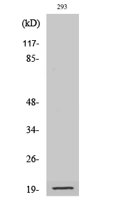
Western Blot analysis of various cells using Cleaved-Bad (D71) AntibodyIHC
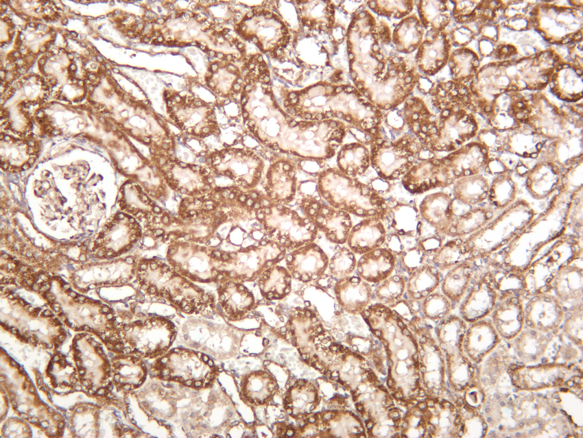
Rat kidney was stained with anti-Bad rabbit antibody.IHC
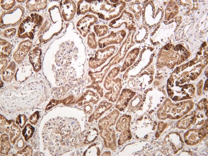
Human kidney was stained with anti-Bad rabbit antibody.IHC
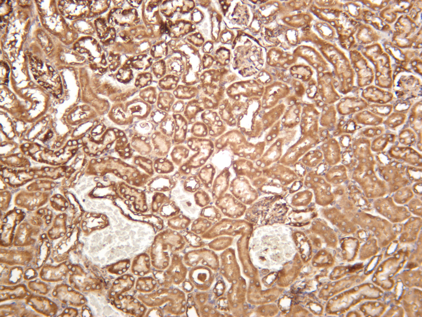
Mouse kidney was stained with anti-Bad rabbit antibody.IF-P
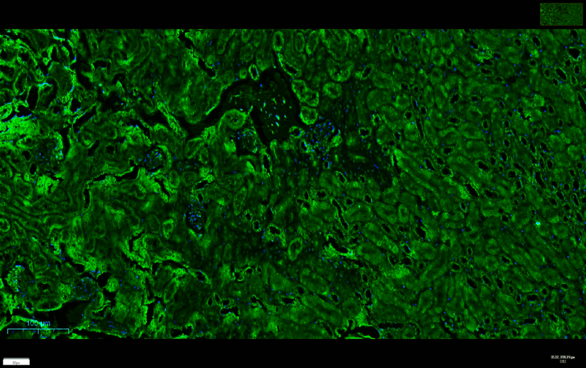
Mouse kidney was stained with anti-Bad rabbit antibody.ICC/IF
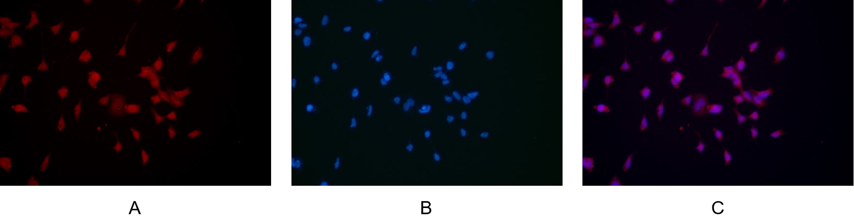
Immunofluorescence analysis of HEK293. Picture A: Bad antibody (red). Picture B: DAPI (blue). Picture C: Merge of A+B.ICC/IF
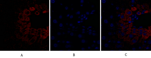
Immunofluorescence analysis of mouse-liver tissue. 1,Bad Antibody (red) was diluted at 1:200 (4℃ overnight). 2, Cy3 labled Secondary antibody was diluted at 1:300 (room temperature, 50min).3, Picture B: DAPI (blue) 10min. Picture A:Target. Picture B: DAPI. Picture C: merge of A+B.| Product Name | Bad Rabbit mAb |
|---|---|
| Antibody Type | Primary Antibodies |
| Immunogen | The specific immunogen used to produce this antibody is proprietary information. |
| Clonality | Monoclonal |
|---|---|
| Isotype | IgG,K |
| Host Species | Rabbit |
| Tested Applications | ICC/IFIF-PIHCWB |
| IHC:1:200-1:1000 WB:1:1000-1:5000 ICC/IF:1:200-1:1000 IF-P:1:200-1:1000 |
|
| Species Reactivity | HumanMouseRat |
| Concentration | 1mg/ml |
| Purification | Protein A |
| Gene Symbol | BAD |
|---|---|
| Gene Synonyms | BBC2 BCL2L8 |
| Gene Full Name | BCL2 associated agonist of cell death |
| Gene Summary | The protein encoded by this gene is a member of the BCL-2 family. BCL-2 family members are known to be regulators of programmed cell death. This protein positively regulates cell apoptosis by forming heterodimers with BCL-xL (B-cell lymphoma-extra large) and BCL-2, and reversing their death repressor activity. Proapoptotic activity of this protein is regulated through its phosphorylation. Protein kinases AKT and MAP kinase, as well as protein phosphatase calcineurin were found to be involved in the regulation of this protein. Alternative splicing of this gene results in two transcript variants which encode the same isoform. [provided by RefSeq, Dec 2019] |
| Alternative Names | BAD BBC6 BCL2L8 Bcl2 antagonist of cell death BAD Bcl-2-binding component 6 Bcl-2-like protein 8 Bcl2-L-8 Bcl-XL/Bcl-2-associated death promoter |
| Molecular Weight(MW) | 18 kDa(Observed band size: 23 kDa) |
| Cellular Localization | Cytoplasm |
| SwissProt ID | Q92934 |
|---|
WB
Various whole cell lysates were separated by 4-20% SDS-PAGE, and the primary antibody was used at 4℃ over night with a 1:2500 dilution . The Dilight 800-conjugated Goat anti-Rabbit antibody was used to detect the antibody.
WB
Various whole cell lysates were separated by 4-20% SDS-PAGE, and the membrane was blotted with anti-Bad antibody. The HRP-conjugated Goat anti-Rabbit IgG (H + L) antibody was used to detect the antibody.
WB
Western Blot analysis of various cells using Cleaved-Bad (D71) Antibody
IHC
Rat kidney was stained with anti-Bad rabbit antibody.
IHC
Human kidney was stained with anti-Bad rabbit antibody.
IHC
Mouse kidney was stained with anti-Bad rabbit antibody.
IF-P
Mouse kidney was stained with anti-Bad rabbit antibody.
ICC/IF
Immunofluorescence analysis of HEK293. Picture A: Bad antibody (red). Picture B: DAPI (blue). Picture C: Merge of A+B.
ICC/IF
Immunofluorescence analysis of mouse-liver tissue. 1,Bad Antibody (red) was diluted at 1:200 (4℃ overnight). 2, Cy3 labled Secondary antibody was diluted at 1:300 (room temperature, 50min).3, Picture B: DAPI (blue) 10min. Picture A:Target. Picture B: DAPI. Picture C: merge of A+B.| Application Notes | IHC:1:200-1:1000 WB:1:1000-1:5000 ICC/IF:1:200-1:1000 IF-P:1:200-1:1000 |
|---|
| Form | Liquid |
|---|---|
| Storage Instructions | -15°C to -25°C/1 year(Do not lower than -25°C). |
| Storage Buffer | PBS, 50% glycerol, 0.05% Proclin 300, 0.05%BSA. |


