IHC
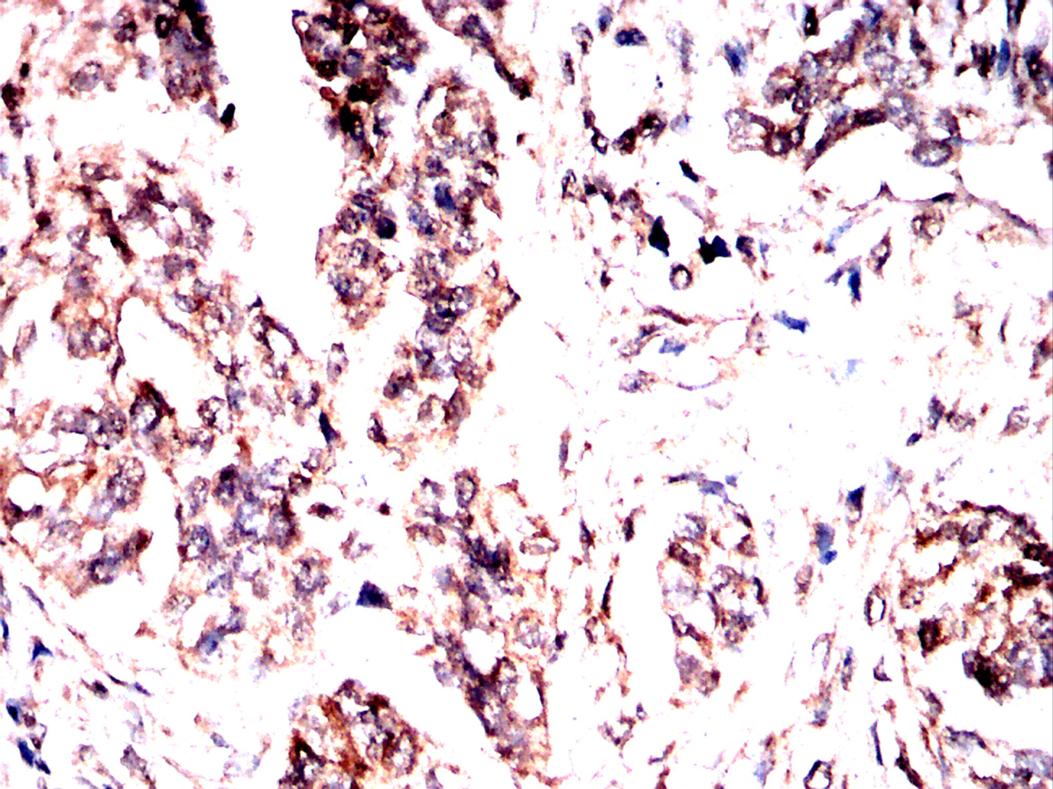
Immunohistochemical analysis of paraffin-embedded Human breast cancer tissues using DDB1 antibody with DAB staining.Pre-treat the sections with heat-mediated antigen retrieval using sodium citrate buffer (pH 6.0) (OM750020) for 2 minutes. Wash the sections with ddH₂O and PBS (OM750003). Block the tissue with 10% non-immune goat serum(OM760028) at room temperature for 30 minutes. Incubate the tissue with the primary antibody diluted at a ratio of 1:1500 at 4°C overnight. At room temperature, dilute the secondary antibody, Goat Anti-Rabbit IgG(H&L)-HRP (OM643487), at a ratio of 1:200 and incubate for one hour. Use DAB(OM760029)as the chromogenic agent. Counterstain the tissue with hematoxylin, and mount the tissue sections with neutral gum.IHC
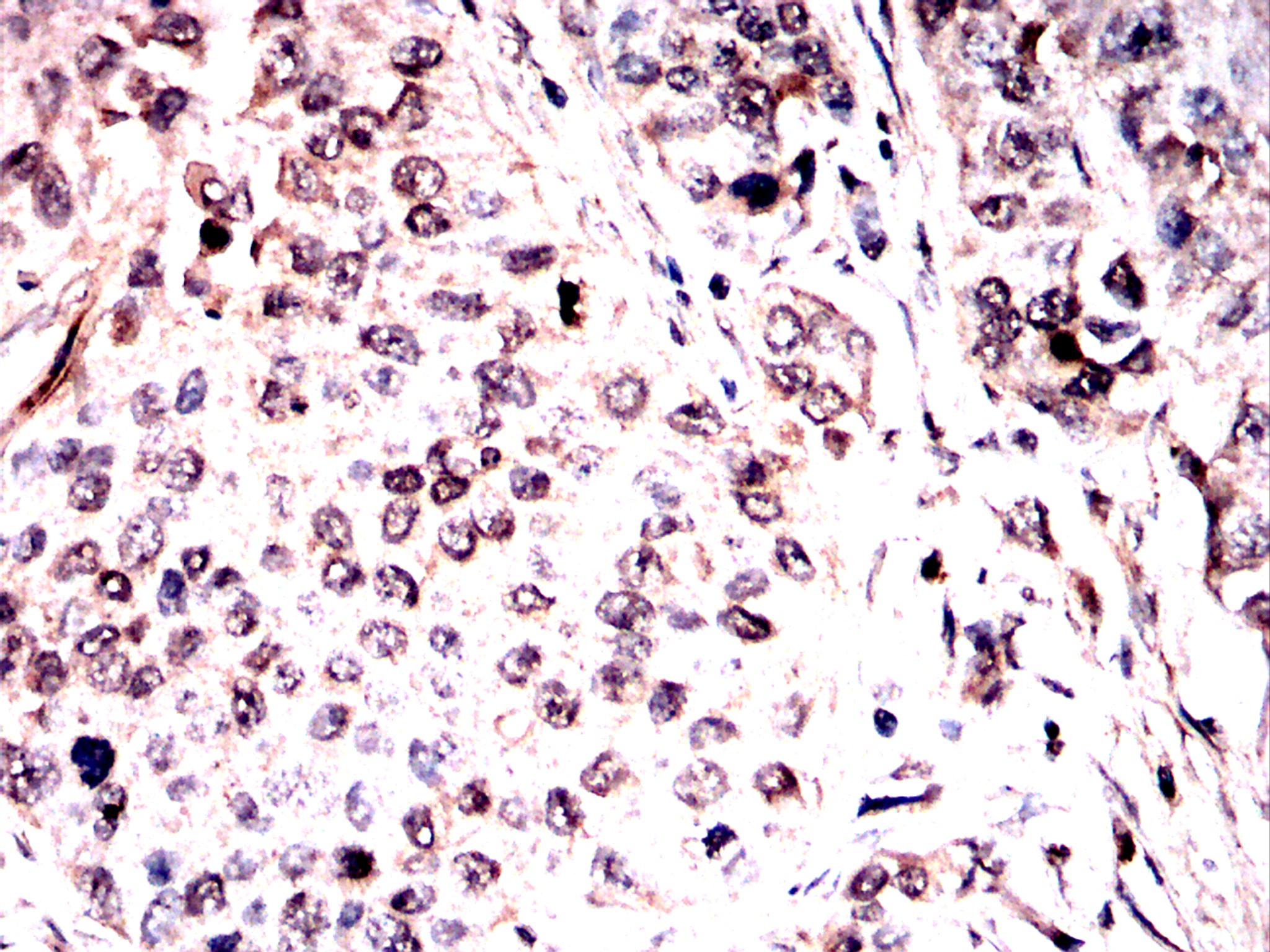
Immunohistochemical analysis of paraffin-embedded Human cervical cancer tissues using DDB1 antibody with DAB staining.Pre-treat the sections with heat-mediated antigen retrieval using sodium citrate buffer (pH 6.0) (OM750020) for 2 minutes. Wash the sections with ddH₂O and PBS (OM750003). Block the tissue with 10% non-immune goat serum(OM760028) at room temperature for 30 minutes. Incubate the tissue with the primary antibody diluted at a ratio of 1:1500 at 4°C overnight. At room temperature, dilute the secondary antibody, Goat Anti-Rabbit IgG(H&L)-HRP (OM643487), at a ratio of 1:200 and incubate for one hour. Use DAB(OM760029)as the chromogenic agent. Counterstain the tissue with hematoxylin, and mount the tissue sections with neutral gum.IHC
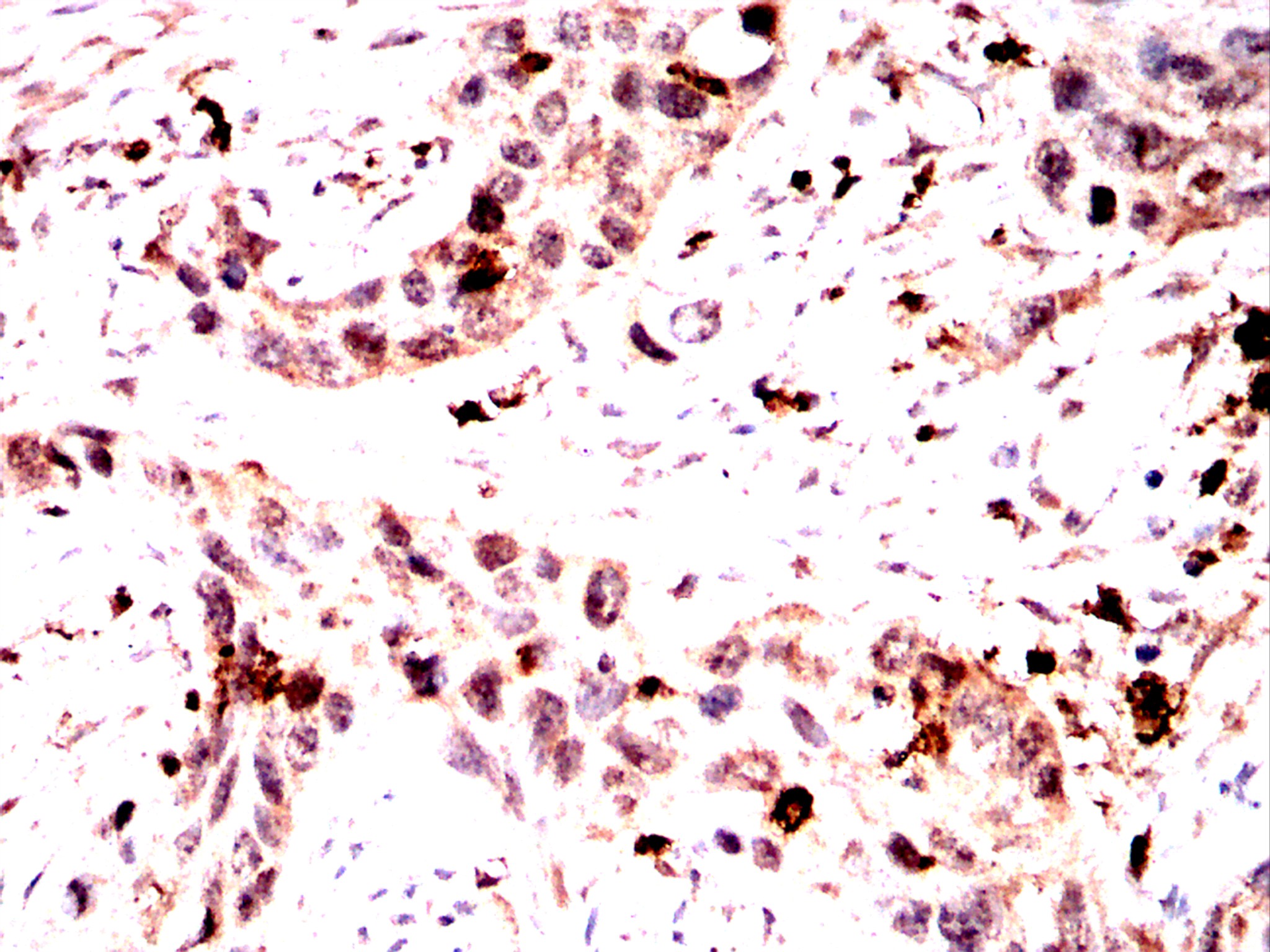
Immunohistochemical analysis of paraffin-embedded Human lung cancer tissues using DDB1 antibody with DAB staining.Pre-treat the sections with heat-mediated antigen retrieval using sodium citrate buffer (pH 6.0) (OM750020) for 2 minutes. Wash the sections with ddH₂O and PBS (OM750003). Block the tissue with 10% non-immune goat serum(OM760028) at room temperature for 30 minutes. Incubate the tissue with the primary antibody diluted at a ratio of 1:1500 at 4°C overnight. At room temperature, dilute the secondary antibody, Goat Anti-Rabbit IgG(H&L)-HRP (OM643487), at a ratio of 1:200 and incubate for one hour. Use DAB(OM760029)as the chromogenic agent. Counterstain the tissue with hematoxylin, and mount the tissue sections with neutral gum.IHC
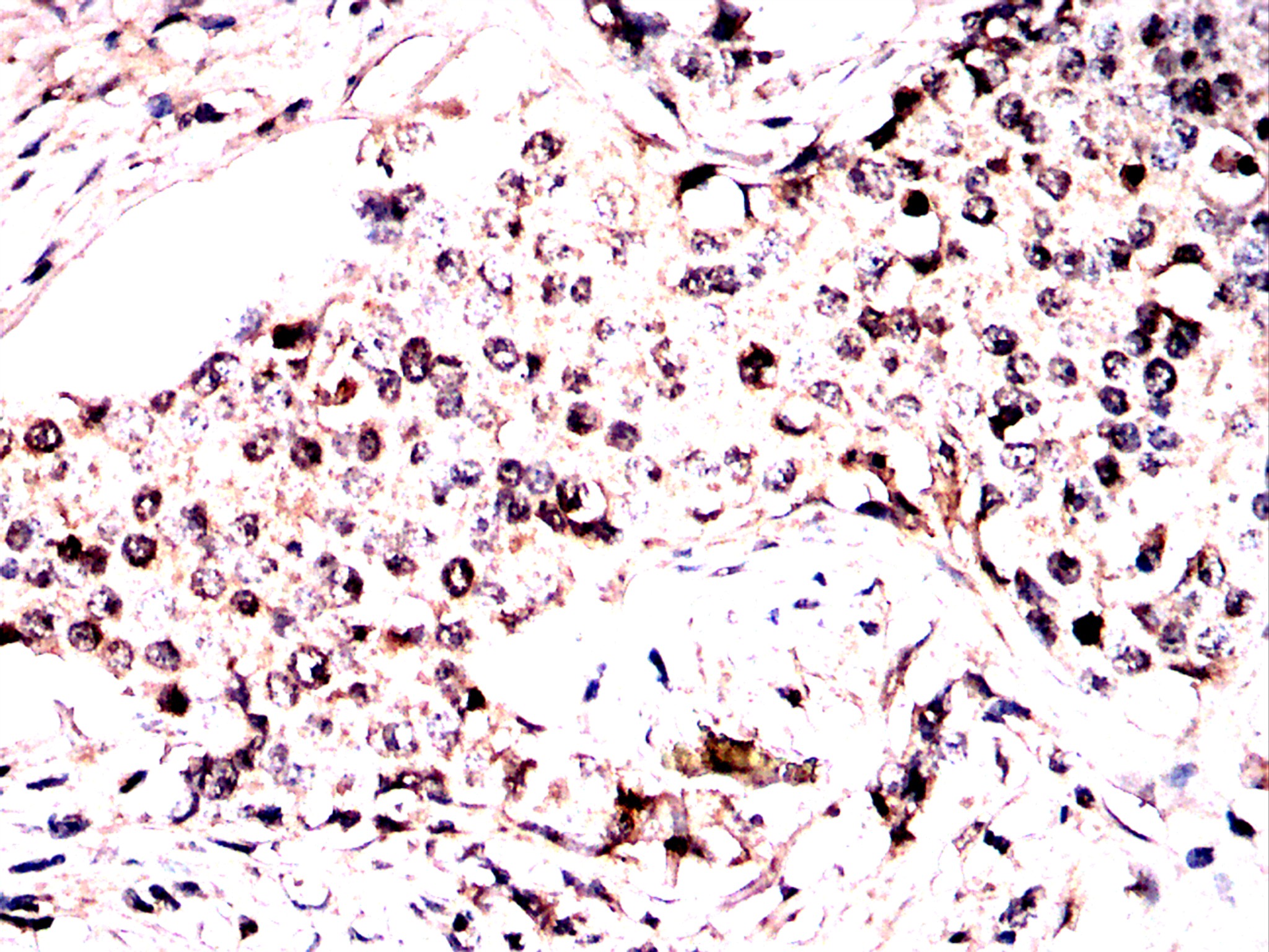
Immunohistochemical analysis of paraffin-embedded Human ovarian cancer tissues using DDB1 antibody with DAB staining.Pre-treat the sections with heat-mediated antigen retrieval using sodium citrate buffer (pH 6.0) (OM750020) for 2 minutes. Wash the sections with ddH₂O and PBS (OM750003). Block the tissue with 10% non-immune goat serum(OM760028) at room temperature for 30 minutes. Incubate the tissue with the primary antibody diluted at a ratio of 1:1500 at 4°C overnight. At room temperature, dilute the secondary antibody, Goat Anti-Rabbit IgG(H&L)-HRP (OM643487), at a ratio of 1:200 and incubate for one hour. Use DAB(OM760029)as the chromogenic agent. Counterstain the tissue with hematoxylin, and mount the tissue sections with neutral gum.WB
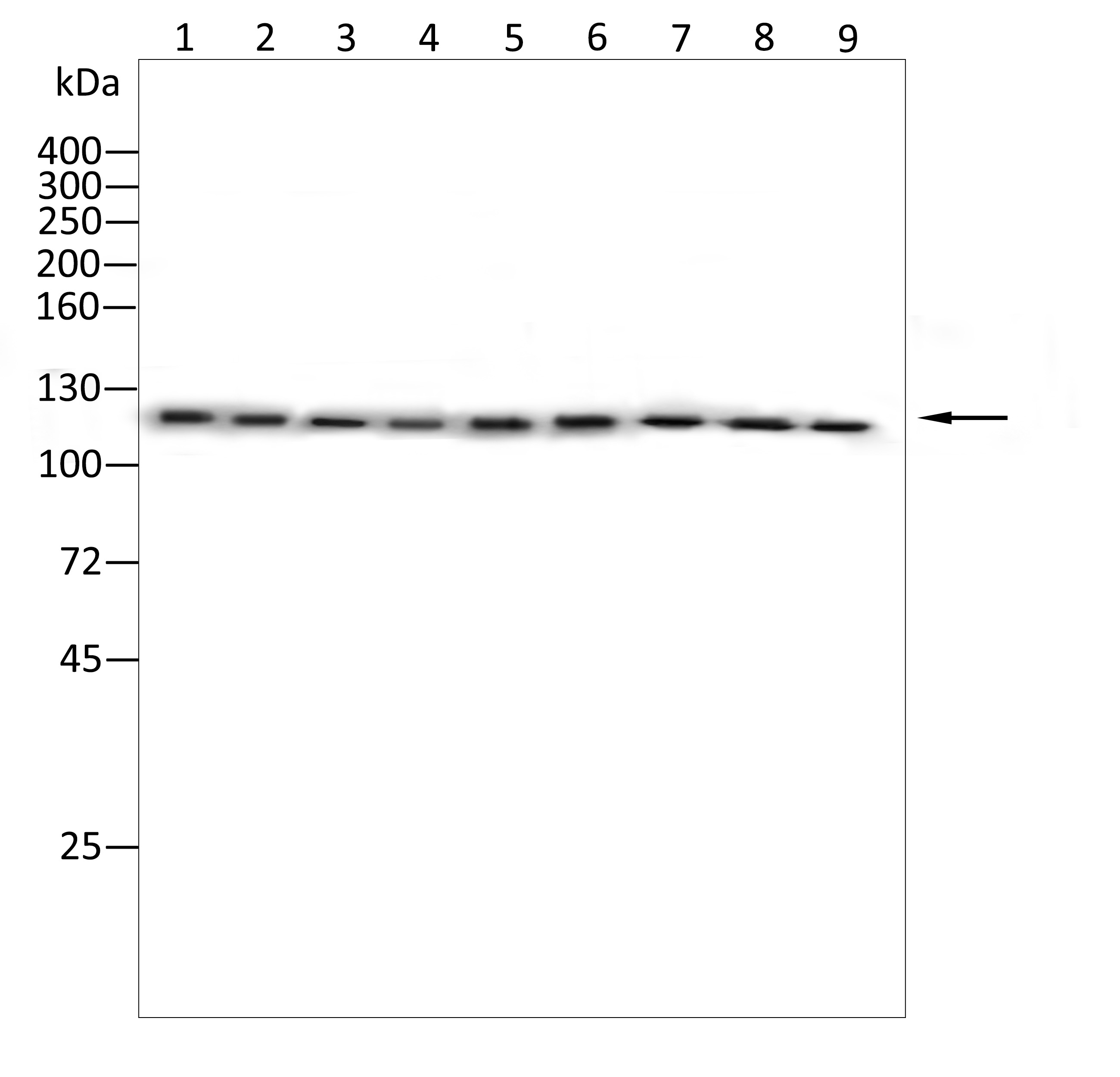
Western blot analysis using DDB1 antibody against Hela(1),A549(2),A431(3),Huvec(4),Cos-7(5),Hek293(6),Mcf-7(7),Jurkat(8),Raji(9) cell . 12% SDS-PAGE gel.Sample loading: 20μg /lane. Transfer the proteins onto a PVDF membrane (OM790003), and block it with TBST (OM750016) plus skimmed milk powder(OM750084) for one hour. Dilute the primary antibody with the antibody diluent (OM750012) at a ratio of 1:2000, and incubate it overnight at 4°C. Wash the membrane three times with TBST (OM750016), 5 minutes each time. At room temperature, dilute the secondary antibody, Goat Anti-Rabbit IgG(H&L)-HRP (OM643487), at a ratio of 1:20000 and incubate for one hour. Wash the membrane three times with TBST (OM750016) again, 5 minutes each time. Use ECL (OM625701) for luminescence.staining time: 60S.ELISA
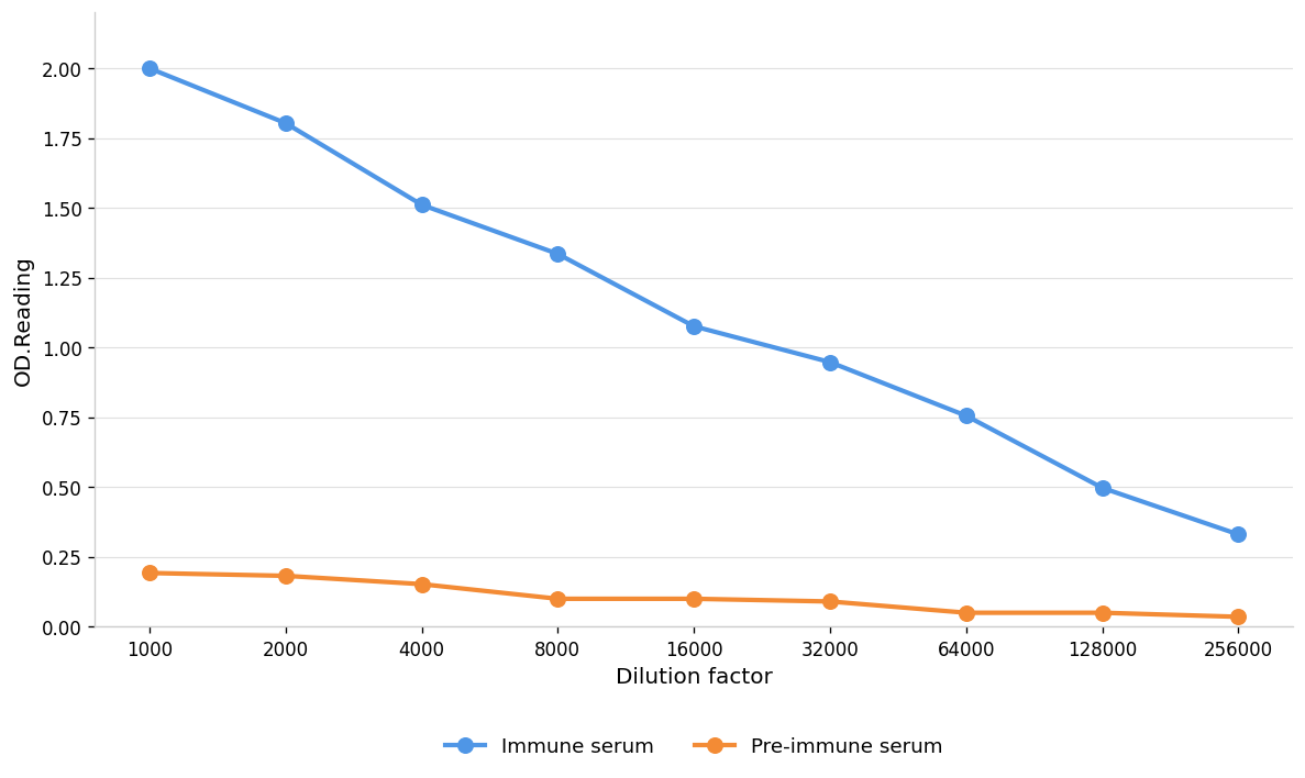
Coat the plate with immunizing antigen.Blue line: Immune serum; Orange line: Pre-immune serum.| Product Name | Anti-DDB1 antibody |
|---|---|
| Antibody Type | Primary Antibodies |
| Immunogen | Polypeptide |
| Clonality | Polyclonal |
|---|---|
| Isotype | IgG |
| Host Species | Rabbit |
| Tested Applications | ELISAIHCWB |
| IHC:1:200-1:2000 WB:1:1000-1:2000 ELISA:1:1000-1:10000 |
|
| Species Reactivity | HumanMouseRat |
| Concentration | 1mg/ml |
| Purification | Protein A |
| Gene Symbol | DDB1 |
|---|---|
| Gene Synonyms | 127kDa DDBA p127-Ddb1 UV-DDB1 WHIKERS XAP1 XPCE XPE XPE-BF |
| Gene Full Name | damage specific DNA binding protein 1 |
| Gene Summary | The protein encoded by this gene is the large subunit (p127) of the heterodimeric DNA damage-binding (DDB) complex while another protein (p48) forms the small subunit. This protein complex functions in nucleotide-excision repair and binds to DNA following UV damage. Defective activity of this complex causes the repair defect in patients with xeroderma pigmentosum complementation group E (XPE) - an autosomal recessive disorder characterized by photosensitivity and early onset of carcinomas. However, it remains for mutation analysis to demonstrate whether the defect in XPE patients is in this gene or the gene encoding the small subunit. In addition, Best vitelliform mascular dystrophy is mapped to the same region as this gene on 11q, but no sequence alternations of this gene are demonstrated in Best disease patients. The protein encoded by this gene also functions as an adaptor molecule for the cullin 4 (CUL4) ubiquitin E3 ligase complex by facilitating the binding of substrates to this complex and the ubiquitination of proteins. [provided by RefSeq, May 2012] |
| Alternative Names | damage-specific DNA binding protein 1 127kDa damage-specific DNA-binding protein DNA repair damaged-DNA recognition protein 1 DDB p127 subunit DNA damage-binding protein a DNA repair protein HBV X-associated protein 1 UV-damaged DNA-binding factor UV-damaged DNA-binding protein 1 UV-DDB 1 XAP-1 xeroderma pigmento xeroderma pigmentosum group E-complementing protein XPE-binding factor |
| Molecular Weight(MW) | 127kDa |
| Source | Rabbit |
| Cellular Localization | Cytoplasm, Nucleus |
| SwissProt ID | Q16531 |
|---|
IHC
Immunohistochemical analysis of paraffin-embedded Human breast cancer tissues using DDB1 antibody with DAB staining.Pre-treat the sections with heat-mediated antigen retrieval using sodium citrate buffer (pH 6.0) (OM750020) for 2 minutes. Wash the sections with ddH₂O and PBS (OM750003). Block the tissue with 10% non-immune goat serum(OM760028) at room temperature for 30 minutes. Incubate the tissue with the primary antibody diluted at a ratio of 1:1500 at 4°C overnight. At room temperature, dilute the secondary antibody, Goat Anti-Rabbit IgG(H&L)-HRP (OM643487), at a ratio of 1:200 and incubate for one hour. Use DAB(OM760029)as the chromogenic agent. Counterstain the tissue with hematoxylin, and mount the tissue sections with neutral gum.
IHC
Immunohistochemical analysis of paraffin-embedded Human cervical cancer tissues using DDB1 antibody with DAB staining.Pre-treat the sections with heat-mediated antigen retrieval using sodium citrate buffer (pH 6.0) (OM750020) for 2 minutes. Wash the sections with ddH₂O and PBS (OM750003). Block the tissue with 10% non-immune goat serum(OM760028) at room temperature for 30 minutes. Incubate the tissue with the primary antibody diluted at a ratio of 1:1500 at 4°C overnight. At room temperature, dilute the secondary antibody, Goat Anti-Rabbit IgG(H&L)-HRP (OM643487), at a ratio of 1:200 and incubate for one hour. Use DAB(OM760029)as the chromogenic agent. Counterstain the tissue with hematoxylin, and mount the tissue sections with neutral gum.
IHC
Immunohistochemical analysis of paraffin-embedded Human lung cancer tissues using DDB1 antibody with DAB staining.Pre-treat the sections with heat-mediated antigen retrieval using sodium citrate buffer (pH 6.0) (OM750020) for 2 minutes. Wash the sections with ddH₂O and PBS (OM750003). Block the tissue with 10% non-immune goat serum(OM760028) at room temperature for 30 minutes. Incubate the tissue with the primary antibody diluted at a ratio of 1:1500 at 4°C overnight. At room temperature, dilute the secondary antibody, Goat Anti-Rabbit IgG(H&L)-HRP (OM643487), at a ratio of 1:200 and incubate for one hour. Use DAB(OM760029)as the chromogenic agent. Counterstain the tissue with hematoxylin, and mount the tissue sections with neutral gum.
IHC
Immunohistochemical analysis of paraffin-embedded Human ovarian cancer tissues using DDB1 antibody with DAB staining.Pre-treat the sections with heat-mediated antigen retrieval using sodium citrate buffer (pH 6.0) (OM750020) for 2 minutes. Wash the sections with ddH₂O and PBS (OM750003). Block the tissue with 10% non-immune goat serum(OM760028) at room temperature for 30 minutes. Incubate the tissue with the primary antibody diluted at a ratio of 1:1500 at 4°C overnight. At room temperature, dilute the secondary antibody, Goat Anti-Rabbit IgG(H&L)-HRP (OM643487), at a ratio of 1:200 and incubate for one hour. Use DAB(OM760029)as the chromogenic agent. Counterstain the tissue with hematoxylin, and mount the tissue sections with neutral gum.
WB
Western blot analysis using DDB1 antibody against Hela(1),A549(2),A431(3),Huvec(4),Cos-7(5),Hek293(6),Mcf-7(7),Jurkat(8),Raji(9) cell . 12% SDS-PAGE gel.Sample loading: 20μg /lane. Transfer the proteins onto a PVDF membrane (OM790003), and block it with TBST (OM750016) plus skimmed milk powder(OM750084) for one hour. Dilute the primary antibody with the antibody diluent (OM750012) at a ratio of 1:2000, and incubate it overnight at 4°C. Wash the membrane three times with TBST (OM750016), 5 minutes each time. At room temperature, dilute the secondary antibody, Goat Anti-Rabbit IgG(H&L)-HRP (OM643487), at a ratio of 1:20000 and incubate for one hour. Wash the membrane three times with TBST (OM750016) again, 5 minutes each time. Use ECL (OM625701) for luminescence.staining time: 60S.
ELISA
Coat the plate with immunizing antigen.Blue line: Immune serum; Orange line: Pre-immune serum.| Application Notes | IHC:1:200-1:2000 WB:1:1000-1:2000 ELISA:1:1000-1:10000 |
|---|
| Form | Liquid |
|---|---|
| Storage Instructions | Shipped at 4°C. Store at +4°C short term (1-2 weeks). Store at -20°C long term. Avoid freeze / thaw cycle. |
| Storage Buffer | Purified antibody in PBS with 0.05% PC300,40% Glycerol. |


