WB
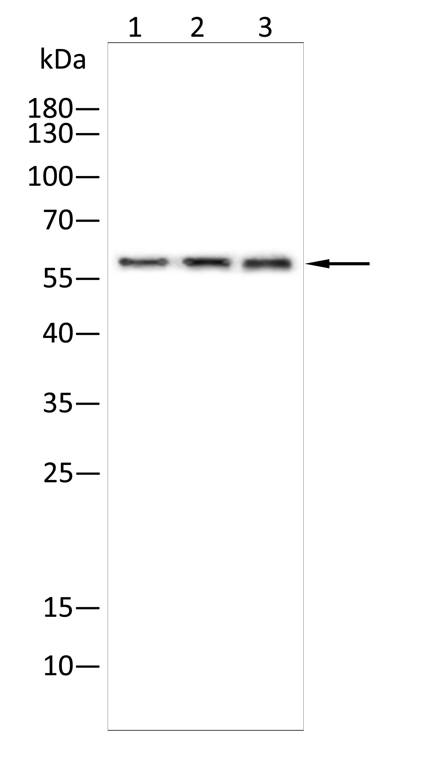
Western blot analysis using UAP1 antibody against K562(1),Pc-12(2),Raji(3) cell . 12% SDS-PAGE gel.Sample loading: 20μg /lane. Transfer the proteins onto a PVDF membrane (OM790003), and block it with TBST (OM750016) plus skimmed milk powder(OM750084) for one hour. Dilute the primary antibody with the antibody diluent (OM750012) at a ratio of 1:2000, and incubate it overnight at 4°C. Wash the membrane three times with TBST (OM750016), 5 minutes each time. At room temperature, dilute the secondary antibody, Goat Anti-Rabbit IgG(H&L)-HRP (OM643487), at a ratio of 1:20000 and incubate for one hour. Wash the membrane three times with TBST (OM750016) again, 5 minutes each time. Use ECL (OM625701) for luminescence.staining time: 60S.WB
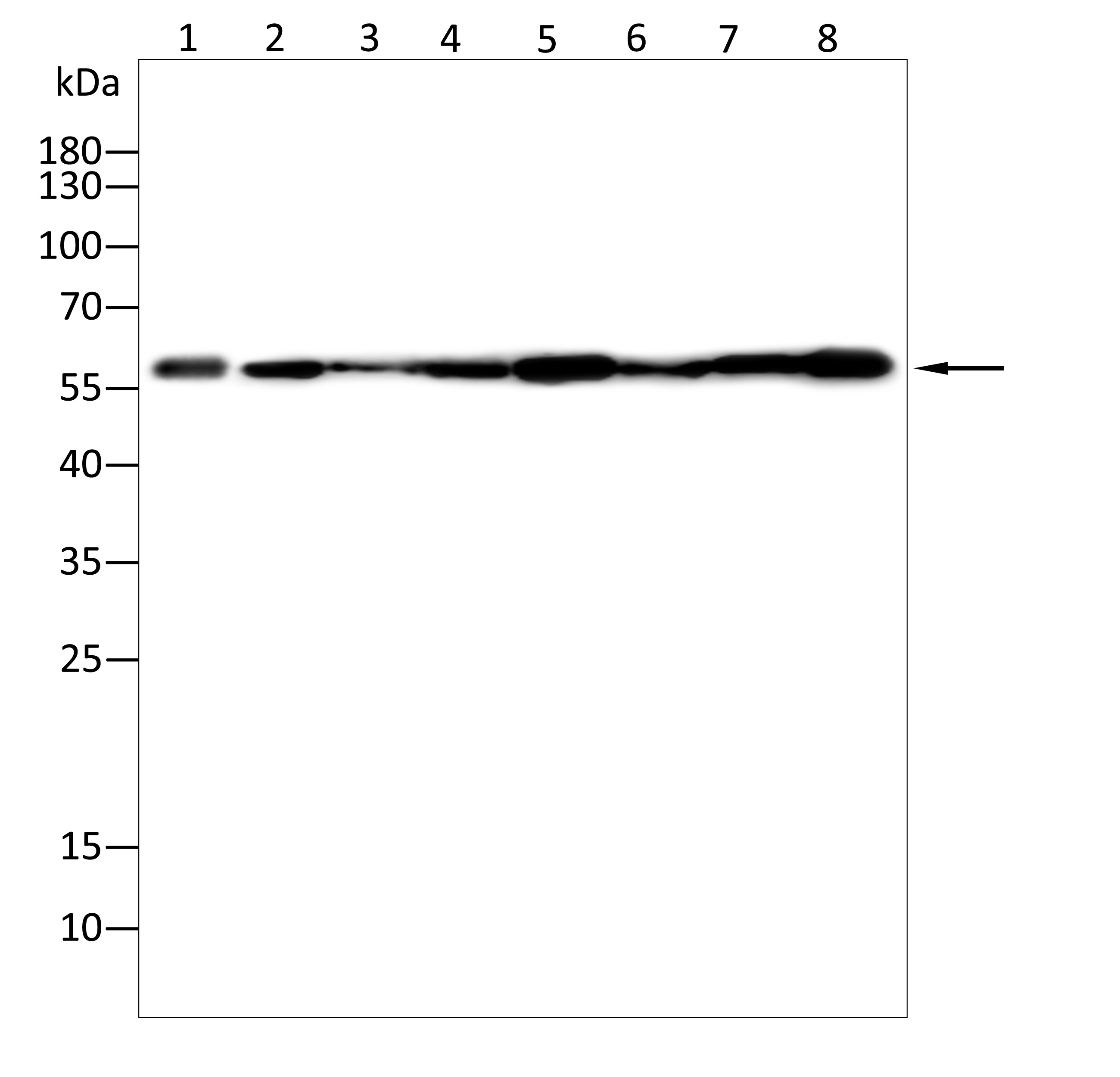
Western blot analysis using UAP1 antibody against Mouse skin tissue(1),Mouse kidney tissue(2),Mouse brain tissue(3),Mouse liver tissue(4),Mouse lung tissue(5),Mouse spleen tissue(6),Mouse stomach tissue(7),Mouse heart tissue(8) . 12% SDS-PAGE gel.Sample loading: 20μg /lane. Transfer the proteins onto a PVDF membrane (OM790003), and block it with TBST (OM750016) plus skimmed milk powder(OM750084) for one hour. Dilute the primary antibody with the antibody diluent (OM750012) at a ratio of 1:2000, and incubate it overnight at 4°C. Wash the membrane three times with TBST (OM750016), 5 minutes each time. At room temperature, dilute the secondary antibody, Goat Anti-Rabbit IgG(H&L)-HRP (OM643487), at a ratio of 1:20000 and incubate for one hour. Wash the membrane three times with TBST (OM750016) again, 5 minutes each time. Use ECL (OM625701) for luminescence.staining time: 60S.ELISA
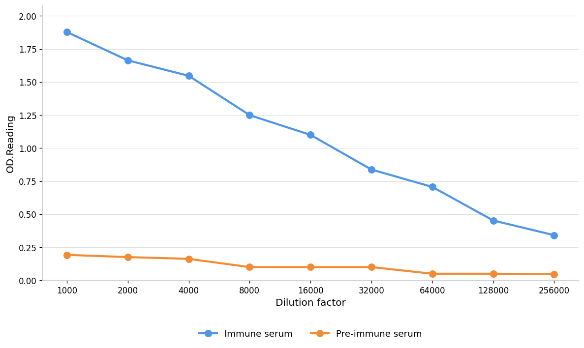
Coat the plate with immunizing antigen.Blue line: Immune serum; Orange line: Pre-immune serum.| Product Name | Anti-UAP1 antibody |
|---|---|
| Antibody Type | Primary Antibodies |
| Immunogen | Polypeptide |
| Clonality | Polyclonal |
|---|---|
| Isotype | IgG |
| Host Species | Rabbit |
| Tested Applications | ELISAWB |
| WB:1:1000-1:2000 ELISA:1:1000-1:10000 |
|
| Species Reactivity | HumanMouseRat |
| Concentration | 1mg/ml |
| Purification | Protein A |
| Gene Symbol | UAP1 |
|---|---|
| Gene Synonyms | AGX AGX-1 AGX-2 AGX1 AGX2 ESTM38 RGD1561967 SPAG2 |
| Gene Full Name | UDP-N-acetylglucosamine pyrophosphorylase 1 |
| Gene Summary | Enables identical protein binding activity and protein serine pyrophosphorylase activity. Involved in antiviral innate immune response and positive regulation of type I interferon production. Located in nucleoplasm and plasma membrane. Is active in cytosol. [provided by Alliance of Genome Resources, Jul 2025] |
| Alternative Names | antigen X protein-pyrophosphorylation enzyme sperm associated antigen 2 testis tissue sperm-binding protein Li 37a UDP-N-acetylgalactosamine pyrophosphorylase UDP-N-acetylglucosamine diphosphorylase 1 UDP-N-acetylhexosamine pyrophosphorylase 1 UDP-N-acteylglucosamine pyrophosphorylase 1 |
| Molecular Weight(MW) | 59kDa |
| Cellular Localization | Cytoplasm |
| SwissProt ID | Q16222 |
|---|
WB
Western blot analysis using UAP1 antibody against K562(1),Pc-12(2),Raji(3) cell . 12% SDS-PAGE gel.Sample loading: 20μg /lane. Transfer the proteins onto a PVDF membrane (OM790003), and block it with TBST (OM750016) plus skimmed milk powder(OM750084) for one hour. Dilute the primary antibody with the antibody diluent (OM750012) at a ratio of 1:2000, and incubate it overnight at 4°C. Wash the membrane three times with TBST (OM750016), 5 minutes each time. At room temperature, dilute the secondary antibody, Goat Anti-Rabbit IgG(H&L)-HRP (OM643487), at a ratio of 1:20000 and incubate for one hour. Wash the membrane three times with TBST (OM750016) again, 5 minutes each time. Use ECL (OM625701) for luminescence.staining time: 60S.
WB
Western blot analysis using UAP1 antibody against Mouse skin tissue(1),Mouse kidney tissue(2),Mouse brain tissue(3),Mouse liver tissue(4),Mouse lung tissue(5),Mouse spleen tissue(6),Mouse stomach tissue(7),Mouse heart tissue(8) . 12% SDS-PAGE gel.Sample loading: 20μg /lane. Transfer the proteins onto a PVDF membrane (OM790003), and block it with TBST (OM750016) plus skimmed milk powder(OM750084) for one hour. Dilute the primary antibody with the antibody diluent (OM750012) at a ratio of 1:2000, and incubate it overnight at 4°C. Wash the membrane three times with TBST (OM750016), 5 minutes each time. At room temperature, dilute the secondary antibody, Goat Anti-Rabbit IgG(H&L)-HRP (OM643487), at a ratio of 1:20000 and incubate for one hour. Wash the membrane three times with TBST (OM750016) again, 5 minutes each time. Use ECL (OM625701) for luminescence.staining time: 60S.
ELISA
Coat the plate with immunizing antigen.Blue line: Immune serum; Orange line: Pre-immune serum.| Application Notes | WB:1:1000-1:2000 ELISA:1:1000-1:10000 |
|---|
| Form | Liquid |
|---|---|
| Storage Instructions | Shipped at 4°C. Store at +4°C short term (1-2 weeks). Store at -20°C long term. Avoid freeze / thaw cycle. |
| Storage Buffer | Purified antibody in PBS with 0.05% PC300,40% Glycerol. |


