WB
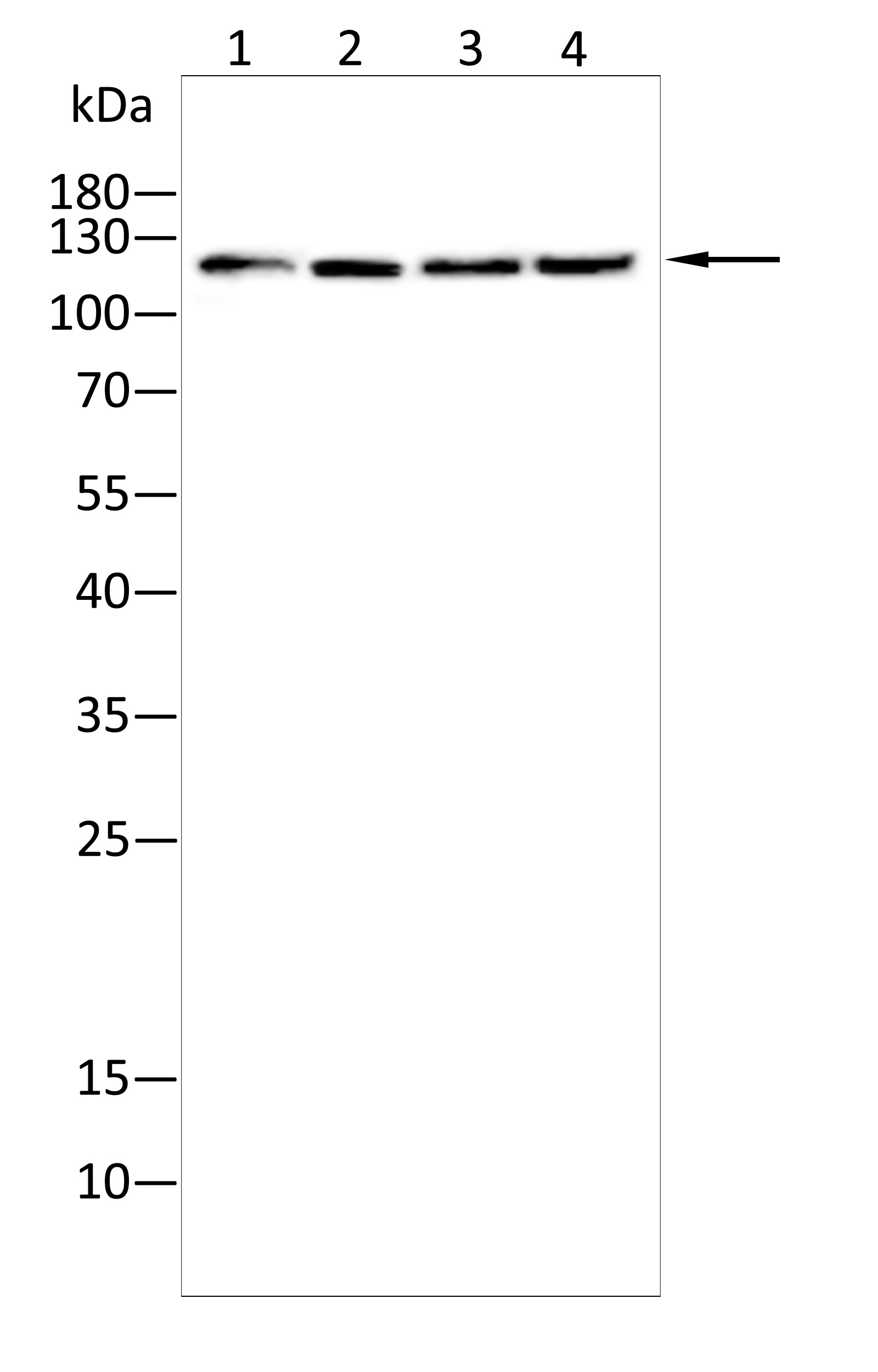
Western blot analysis using SEMA6D antibody against Huvec(1),Mcf-7(2),Pc-12(3) cell,Rat intestine tissue(4) . 12% SDS-PAGE gel.Sample loading: 20μg /lane. Transfer the proteins onto a PVDF membrane (OM790003), and block it with TBST (OM750016) plus skimmed milk powder(OM750084) for one hour. Dilute the primary antibody with the antibody diluent (OM750012) at a ratio of 1:1000, and incubate it overnight at 4°C. Wash the membrane three times with TBST (OM750016), 5 minutes each time. At room temperature, dilute the secondary antibody, Goat Anti-Rabbit IgG(H&L)-HRP (OM643487), at a ratio of 1:20000 and incubate for one hour. Wash the membrane three times with TBST (OM750016) again, 5 minutes each time. Use ECL (OM625701) for luminescence.staining time: 60S.ELISA
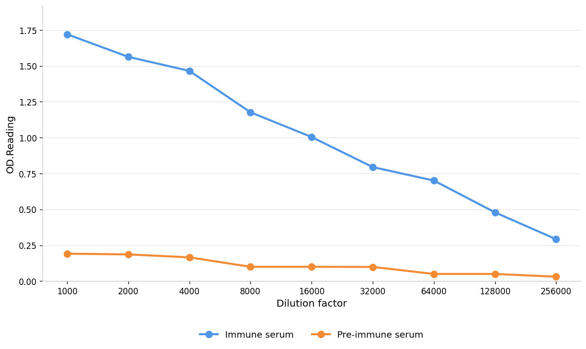
Coat the plate with immunizing antigen.Blue line: Immune serum; Orange line: Pre-immune serum.ICC/IF
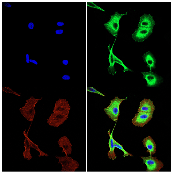
Immunofluorescence analysis of A549 cells using SEMA6Dantibody (green). Blue: DAPI fluorescent DNA dye. Red: Actin filaments have been labeled with Omnimabs® 594-Phalloidin.Cells are fixed in 4% paraformaldehyde at room temperature for 15 minutes. Then, they are permeabilized with a PBS (OM750003) solution containing 0.1% Triton X-100(OM750021) at room temperature for 15 minutes. Subsequently, the cells are blocked with 10% non - immune goat serum(OM760028) at room temperature for 1 hour.The cells are incubated overnight at 4°C with the primary antibody diluted 1:100 in PBS. The secondary antibody, Omnimabs® 488 Goat Ant-Rabbit IgG(H&L) (Green,OM643486), is diluted at a ratio of 1:400 and incubated with the cells for 1 hour.Nuclear DNA is labeled with DAPI (Blue,OM643160). F-actin is stained with Omnimabs® 594-Phalloidin (Red,OM750007) diluted 1:100 for 30 minutes.| Product Name | Anti-SEMA6D antibody |
|---|---|
| Antibody Type | Primary Antibodies |
| Immunogen | Polypeptide |
| Clonality | Polyclonal |
|---|---|
| Isotype | IgG |
| Host Species | Rabbit |
| Tested Applications | ELISAICC/IFWB |
| WB:1:1000-1:2000 ICC/IF:1:100-1:500 ELISA:1:1000-1:10000 |
|
| Species Reactivity | HumanMouseRat |
| Concentration | 1mg/ml |
| Purification | Protein A |
| Gene Symbol | SEMA6D |
|---|---|
| Gene Synonyms | 1110067B02Rik D330011G23 mKIAA1479 |
| Gene Full Name | semaphorin 6D |
| Gene Summary | Semaphorins are a large family, including both secreted and membrane associated proteins, many of which have been implicated as inhibitors or chemorepellents in axon pathfinding, fasciculation and branching, and target selection. All semaphorins possess a semaphorin (Sema) domain and a PSI domain (found in plexins, semaphorins and integrins) in the N-terminal extracellular portion. Additional sequence motifs C-terminal to the semaphorin domain allow classification into distinct subfamilies. Results demonstrate that transmembrane semaphorins, like the secreted ones, can act as repulsive axon guidance cues. This gene encodes a class 6 vertebrate transmembrane semaphorin that demonstrates alternative splicing. Several transcript variants have been identified and expression of the distinct encoded isoforms is thought to be regulated in a tissue- and development-dependent manner. [provided by RefSeq, Nov 2010] |
| Alternative Names | axon guidance family member FLJ11598 KIAA1479FLJ11598 sema domain transmembrane domain (TM) and cytoplasmic domain (semaphorin) 6D |
| Molecular Weight(MW) | 119kDa |
| Source | Rabbit |
| Cellular Localization | Cell membrane;Cytoplasm |
| SwissProt ID | Q8NFY4 |
|---|
WB
Western blot analysis using SEMA6D antibody against Huvec(1),Mcf-7(2),Pc-12(3) cell,Rat intestine tissue(4) . 12% SDS-PAGE gel.Sample loading: 20μg /lane. Transfer the proteins onto a PVDF membrane (OM790003), and block it with TBST (OM750016) plus skimmed milk powder(OM750084) for one hour. Dilute the primary antibody with the antibody diluent (OM750012) at a ratio of 1:1000, and incubate it overnight at 4°C. Wash the membrane three times with TBST (OM750016), 5 minutes each time. At room temperature, dilute the secondary antibody, Goat Anti-Rabbit IgG(H&L)-HRP (OM643487), at a ratio of 1:20000 and incubate for one hour. Wash the membrane three times with TBST (OM750016) again, 5 minutes each time. Use ECL (OM625701) for luminescence.staining time: 60S.
ELISA
Coat the plate with immunizing antigen.Blue line: Immune serum; Orange line: Pre-immune serum.
ICC/IF
Immunofluorescence analysis of A549 cells using SEMA6Dantibody (green). Blue: DAPI fluorescent DNA dye. Red: Actin filaments have been labeled with Omnimabs® 594-Phalloidin.Cells are fixed in 4% paraformaldehyde at room temperature for 15 minutes. Then, they are permeabilized with a PBS (OM750003) solution containing 0.1% Triton X-100(OM750021) at room temperature for 15 minutes. Subsequently, the cells are blocked with 10% non - immune goat serum(OM760028) at room temperature for 1 hour.The cells are incubated overnight at 4°C with the primary antibody diluted 1:100 in PBS. The secondary antibody, Omnimabs® 488 Goat Ant-Rabbit IgG(H&L) (Green,OM643486), is diluted at a ratio of 1:400 and incubated with the cells for 1 hour.Nuclear DNA is labeled with DAPI (Blue,OM643160). F-actin is stained with Omnimabs® 594-Phalloidin (Red,OM750007) diluted 1:100 for 30 minutes.| Application Notes | WB:1:1000-1:2000 ICC/IF:1:100-1:500 ELISA:1:1000-1:10000 |
|---|
| Form | Liquid |
|---|---|
| Storage Instructions | Shipped at 4°C. Store at +4°C short term (1-2 weeks). Store at -20°C long term. Avoid freeze / thaw cycle. |
| Storage Buffer | Purified antibody in PBS with 0.05% PC300,40% Glycerol. |


