ELISA
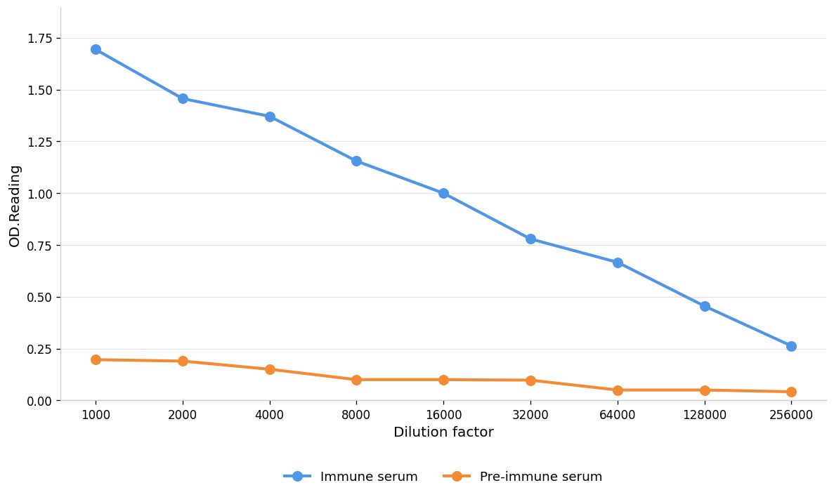
Coat the plate with immunizing antigen.Blue line: Immune serum; Orange line: Pre-immune serum.WB
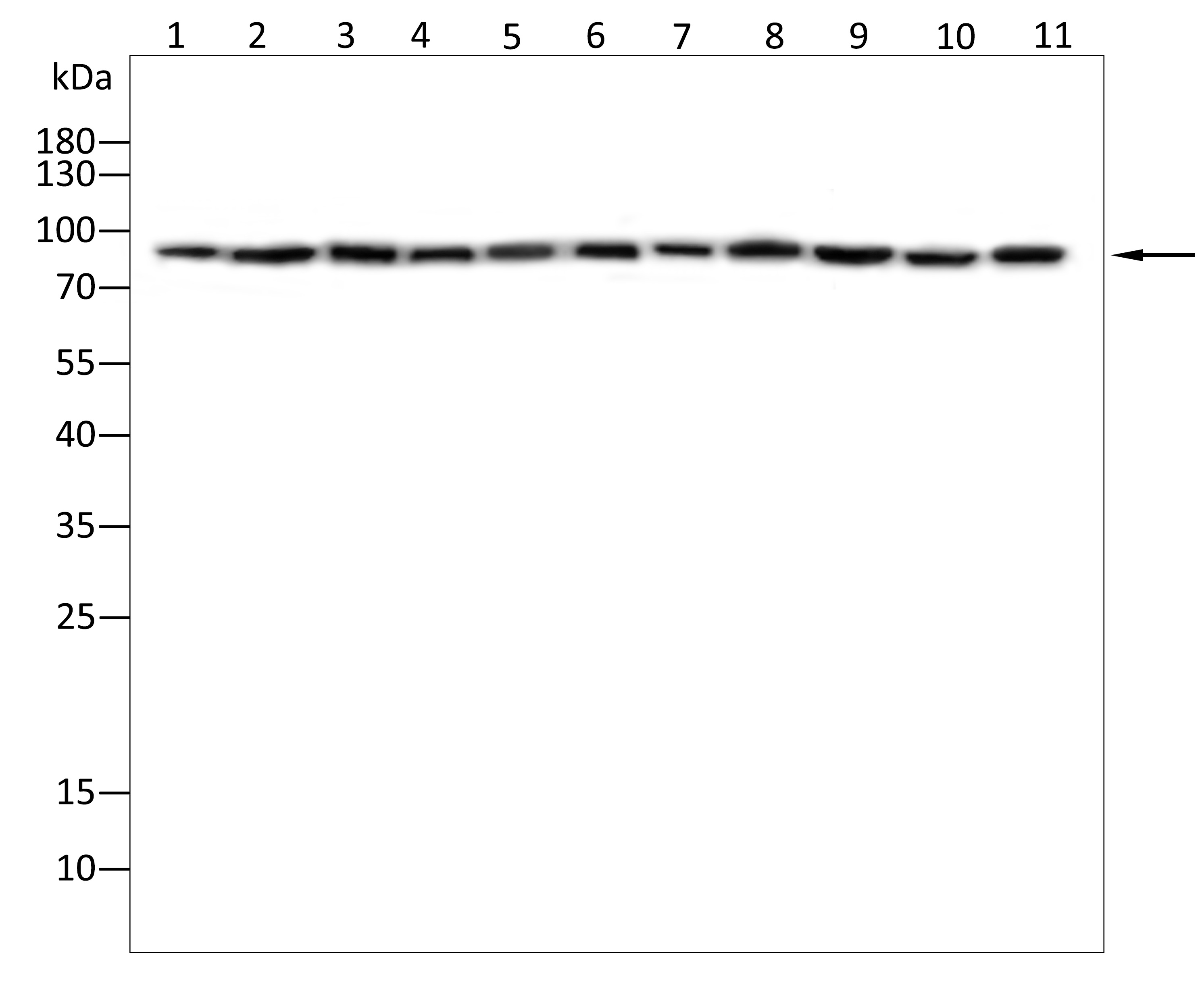
Western blot analysis using ASAP2 antibody against Hela(1),Cos-7(2),K562(3),Hepg2(4),THP-1(5),A549(6),Nih3t3(7),Jurkat(8),C2C12(9),Huvec(10),A431(11)cell . 8% SDS-PAGE gel.Sample loading: 20μg /lane. Transfer the proteins onto a PVDF membrane (OM790003), and block it with TBST (OM750016) plus skimmed milk powder(OM750084) for one hour. Dilute the primary antibody with the antibody diluent (OM750012) at a ratio of 1:1000, and incubate it overnight at 4°C. Wash the membrane three times with TBST (OM750016), 5 minutes each time. At room temperature, dilute the secondary antibody, Goat Anti-Rabbit IgG(H&L)-HRP (OM643487), at a ratio of 1:20000 and incubate for one hour. Wash the membrane three times with TBST (OM750016) again, 5 minutes each time. Use ECL (OM625701) for luminescence.staining time: 60S.WB
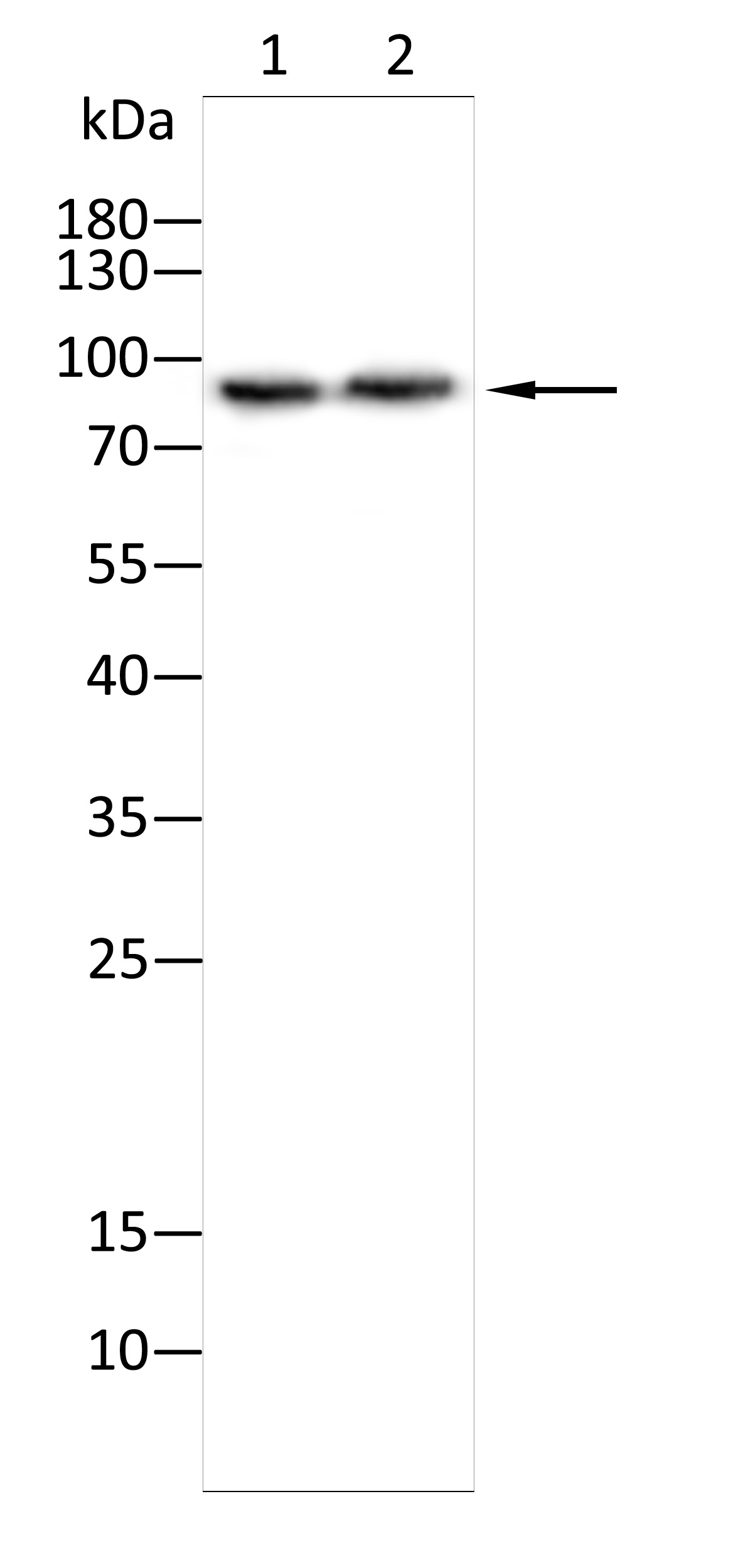
Western blot analysis using ASAP2 antibody against Mouse stomach tissue(1),Rat kidney tissue(2). 8% SDS-PAGE gel.Sample loading: 20μg /lane. Transfer the proteins onto a PVDF membrane (OM790003), and block it with TBST (OM750016) plus skimmed milk powder(OM750084) for one hour. Dilute the primary antibody with the antibody diluent (OM750012) at a ratio of 1:1000, and incubate it overnight at 4°C. Wash the membrane three times with TBST (OM750016), 5 minutes each time. At room temperature, dilute the secondary antibody, Goat Anti-Rabbit IgG(H&L)-HRP (OM643487), at a ratio of 1:20000 and incubate for one hour. Wash the membrane three times with TBST (OM750016) again, 5 minutes each time. Use ECL (OM625701) for luminescence.staining time: 60S.ICC/IF
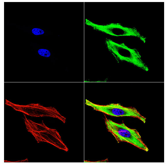
Immunofluorescence analysis of Hela cells using ASAP2 antibody (green). Blue: DAPI fluorescent DNA dye. Red: Actin filaments have been labeled with Omnimabs® 594-Phalloidin.Cells are fixed in 4% paraformaldehyde at room temperature for 15 minutes. Then, they are permeabilized with a PBS (OM750003) solution containing 0.1% Triton X-100(OM750021) at room temperature for 15 minutes. Subsequently, the cells are blocked with 10% non - immune goat serum(OM760028) at room temperature for 1 hour.The cells are incubated overnight at 4°C with the primary antibody diluted 1:100 in PBS. The secondary antibody, Omnimabs® 488 Goat Ant-Rabbit IgG(H&L) (Green,OM643486), is diluted at a ratio of 1:400 and incubated with the cells for 1 hour.Nuclear DNA is labeled with DAPI (Blue,OM643160). F-actin is stained with Omnimabs® 594-Phalloidin (Red,OM750007) diluted 1:100 for 30 minutes.| Product Name | Anti-ASAP2 antibody |
|---|---|
| Antibody Type | Primary Antibodies |
| Immunogen | Polypeptide |
| Clonality | Polyclonal |
|---|---|
| Isotype | IgG |
| Host Species | Rabbit |
| Tested Applications | ELISAICC/IFWB |
| WB:1:1000-1:2000 ICC/IF:1:100-1:500 ELISA:1:1000-1:10000 |
|
| Species Reactivity | HumanMouseRat |
| Concentration | 1mg/ml |
| Purification | Protein A |
| Gene Symbol | ASAP2 |
|---|---|
| Gene Synonyms | 6530401G17Rik AMAP2 CENTB3 DDEF2 Gm1523 Gm592 PAG3 PAP Pap-alpha SHAG1 |
| Gene Full Name | ArfGAP with SH3 domain, ankyrin repeat and PH domain 2 |
| Gene Summary | This gene encodes a multidomain protein containing an N-terminal alpha-helical region with a coiled-coil motif, followed by a pleckstrin homology (PH) domain, an Arf-GAP domain, an ankyrin homology region, a proline-rich region, and a C-terminal Src homology 3 (SH3) domain. The protein localizes in the Golgi apparatus and at the plasma membrane, where it colocalizes with protein tyrosine kinase 2-beta (PYK2). The encoded protein forms a stable complex with PYK2 in vivo. This interaction appears to be mediated by binding of its SH3 domain to the C-terminal proline-rich domain of PYK2. The encoded protein is tyrosine phosphorylated by activated PYK2. It has catalytic activity for class I and II ArfGAPs in vitro, and can bind the class III Arf ARF6 without immediate GAP activity. The encoded protein is believed to function as an ARF GAP that controls ARF-mediated vesicle budding when recruited to Golgi membranes. In addition, it functions as a substrate and downstream target for PYK2 and SRC, a pathway that may be involved in the regulation of vesicular transport. Multiple transcript variants encoding different isoforms have been found for this gene. [provided by RefSeq, Sep 2008] |
| Alternative Names | centaurin beta 3 development and differentiation enhancing factor 2 development and differentiation-enhancing factor 2 KIAA0400 PAPbeta paxillin-associated protein with ARF GAP activity 3 PYK2 C terminus-associated protein pyk2 C-terminus-associated protein |
| Molecular Weight(MW) | 111kDa(observed:90kDa) |
| Source | Rabbit |
| Cellular Localization | Cytoplasm;Golgi apparatus, Golgi stack membrane;Cell membrane |
| SwissProt ID | O43150 |
|---|
ELISA
Coat the plate with immunizing antigen.Blue line: Immune serum; Orange line: Pre-immune serum.
WB
Western blot analysis using ASAP2 antibody against Hela(1),Cos-7(2),K562(3),Hepg2(4),THP-1(5),A549(6),Nih3t3(7),Jurkat(8),C2C12(9),Huvec(10),A431(11)cell . 8% SDS-PAGE gel.Sample loading: 20μg /lane. Transfer the proteins onto a PVDF membrane (OM790003), and block it with TBST (OM750016) plus skimmed milk powder(OM750084) for one hour. Dilute the primary antibody with the antibody diluent (OM750012) at a ratio of 1:1000, and incubate it overnight at 4°C. Wash the membrane three times with TBST (OM750016), 5 minutes each time. At room temperature, dilute the secondary antibody, Goat Anti-Rabbit IgG(H&L)-HRP (OM643487), at a ratio of 1:20000 and incubate for one hour. Wash the membrane three times with TBST (OM750016) again, 5 minutes each time. Use ECL (OM625701) for luminescence.staining time: 60S.
WB
Western blot analysis using ASAP2 antibody against Mouse stomach tissue(1),Rat kidney tissue(2). 8% SDS-PAGE gel.Sample loading: 20μg /lane. Transfer the proteins onto a PVDF membrane (OM790003), and block it with TBST (OM750016) plus skimmed milk powder(OM750084) for one hour. Dilute the primary antibody with the antibody diluent (OM750012) at a ratio of 1:1000, and incubate it overnight at 4°C. Wash the membrane three times with TBST (OM750016), 5 minutes each time. At room temperature, dilute the secondary antibody, Goat Anti-Rabbit IgG(H&L)-HRP (OM643487), at a ratio of 1:20000 and incubate for one hour. Wash the membrane three times with TBST (OM750016) again, 5 minutes each time. Use ECL (OM625701) for luminescence.staining time: 60S.
ICC/IF
Immunofluorescence analysis of Hela cells using ASAP2 antibody (green). Blue: DAPI fluorescent DNA dye. Red: Actin filaments have been labeled with Omnimabs® 594-Phalloidin.Cells are fixed in 4% paraformaldehyde at room temperature for 15 minutes. Then, they are permeabilized with a PBS (OM750003) solution containing 0.1% Triton X-100(OM750021) at room temperature for 15 minutes. Subsequently, the cells are blocked with 10% non - immune goat serum(OM760028) at room temperature for 1 hour.The cells are incubated overnight at 4°C with the primary antibody diluted 1:100 in PBS. The secondary antibody, Omnimabs® 488 Goat Ant-Rabbit IgG(H&L) (Green,OM643486), is diluted at a ratio of 1:400 and incubated with the cells for 1 hour.Nuclear DNA is labeled with DAPI (Blue,OM643160). F-actin is stained with Omnimabs® 594-Phalloidin (Red,OM750007) diluted 1:100 for 30 minutes.| Application Notes | WB:1:1000-1:2000 ICC/IF:1:100-1:500 ELISA:1:1000-1:10000 |
|---|
| Form | Liquid |
|---|---|
| Storage Instructions | Shipped at 4°C. Store at +4°C short term (1-2 weeks). Store at -20°C long term. Avoid freeze / thaw cycle. |
| Storage Buffer | Purified antibody in PBS with 0.05% PC300,40% Glycerol. |


