WB
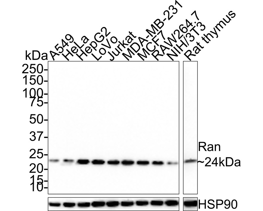
Western blot analysis of Ran on different lysates with Rabbit anti-Ran antibody at 1/1,000 dilution. Lane 1: A549 cell lysate (15 µg/Lane), Lane 2: HeLa cell lysate (15 µg/Lane), Lane 3: HepG2 cell lysate (15 µg/Lane), Lane 4: LoVo cell lysate (15 µg/Lane), Lane 5: Jurkat cell lysate (15 µg/Lane), Lane 6: MDA-MB-231 cell lysate (15 µg/Lane), Lane 7: MCF7 cell lysate (15 µg/Lane), Lane 8: RAW264.7 cell lysate (15 µg/Lane), Lane 9: NIH/3T3 cell lysate (15 µg/Lane), Lane 10: Rat thymus tissue lysate (30 µg/Lane). Exposure time: 40 seconds; 4-20% SDS-PAGE gel. Proteins were transferred to a PVDF membrane and blocked with 5% NFDM/TBST for 1 hour at room temperature. The primary antibody at 1/1,000 dilution was used in 5% NFDM/TBST at room temperature for 2 hours. Goat Anti-Rabbit IgG - HRP Secondary Antibody at 1:100,000 dilution was used for 1 hour at room temperature.FC
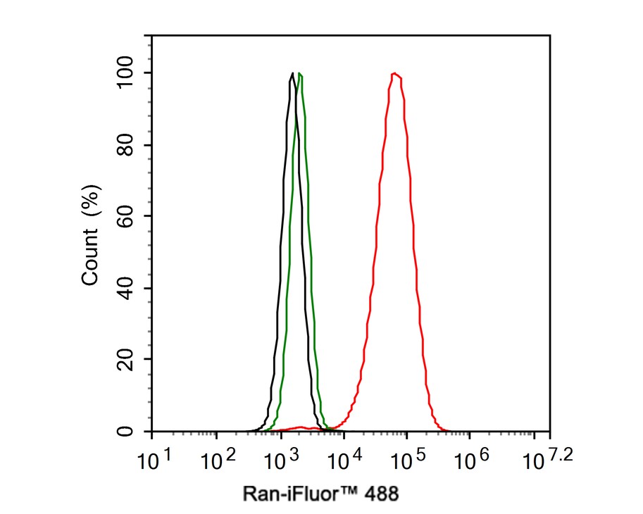
Flow cytometric analysis of HeLa cells labeling Ran. Cells were fixed and permeabilized. Then stained with the primary antibody (1ug/ml) (red) compared with Rabbit IgG Isotype Control (green). After incubation of the primary antibody at +4℃ for an hour, the cells were stained with a 488 conjugate-Goat anti-Rabbit IgG Secondary antibody at 1/1,000 dilution for 30 minutes at +4℃. Unlabelled sample was used as a control (cells without incubation with primary antibody; black).| Product Name | Ran Recombinant Rabbit Monoclonal Antibody |
|---|---|
| Antibody Type | Primary Antibodies |
| Immunogen | Synthetic peptide within Human Ran aa 167-216 / 216. |
| Clonality | Monoclonal |
|---|---|
| Isotype | IgG |
| Host Species | Rabbit |
| Tested Applications | FCWB |
| WB:1:1000 FC:1:500-1:1000 |
|
| Species Reactivity | HumanMouseRat |
| Concentration | 1mg/ml |
| Purification | Protein A |
| Gene Symbol | RAN |
|---|---|
| Gene Synonyms | TC4 Gsp1 ARA24 |
| Gene Full Name | RAN, member RAS oncogene family |
| Gene Summary | RAN (ras-related nuclear protein) is a small GTP binding protein belonging to the RAS superfamily that is essential for the translocation of RNA and proteins through the nuclear pore complex. The RAN protein is also involved in control of DNA synthesis and cell cycle progression. Nuclear localization of RAN requires the presence of regulator of chromosome condensation 1 (RCC1). Mutations in RAN disrupt DNA synthesis. Because of its many functions, it is likely that RAN interacts with several other proteins. RAN regulates formation and organization of the microtubule network independently of its role in the nucleus-cytosol exchange of macromolecules. RAN could be a key signaling molecule regulating microtubule polymerization during mitosis. RCC1 generates a high local concentration of RAN-GTP around chromatin which, in turn, induces the local nucleation of microtubules. RAN is an androgen receptor (AR) coactivator that binds differentially with different lengths of polyglutamine within the androgen receptor. Polyglutamine repeat expansion in the AR is linked to Kennedy's disease (X-linked spinal and bulbar muscular atrophy). RAN coactivation of the AR diminishes with polyglutamine expansion within the AR, and this weak coactivation may lead to partial androgen insensitivity during the development of Kennedy's disease. [provided by RefSeq, Jul 2008] |
| Molecular Weight(MW) | 24 kDa |
| Cellular Localization | Nucleus, Nucleus envelope, Cytoplasm, cytosol, Melanosome. |
WB
Western blot analysis of Ran on different lysates with Rabbit anti-Ran antibody at 1/1,000 dilution. Lane 1: A549 cell lysate (15 µg/Lane), Lane 2: HeLa cell lysate (15 µg/Lane), Lane 3: HepG2 cell lysate (15 µg/Lane), Lane 4: LoVo cell lysate (15 µg/Lane), Lane 5: Jurkat cell lysate (15 µg/Lane), Lane 6: MDA-MB-231 cell lysate (15 µg/Lane), Lane 7: MCF7 cell lysate (15 µg/Lane), Lane 8: RAW264.7 cell lysate (15 µg/Lane), Lane 9: NIH/3T3 cell lysate (15 µg/Lane), Lane 10: Rat thymus tissue lysate (30 µg/Lane). Exposure time: 40 seconds; 4-20% SDS-PAGE gel. Proteins were transferred to a PVDF membrane and blocked with 5% NFDM/TBST for 1 hour at room temperature. The primary antibody at 1/1,000 dilution was used in 5% NFDM/TBST at room temperature for 2 hours. Goat Anti-Rabbit IgG - HRP Secondary Antibody at 1:100,000 dilution was used for 1 hour at room temperature.
FC
Flow cytometric analysis of HeLa cells labeling Ran. Cells were fixed and permeabilized. Then stained with the primary antibody (1ug/ml) (red) compared with Rabbit IgG Isotype Control (green). After incubation of the primary antibody at +4℃ for an hour, the cells were stained with a 488 conjugate-Goat anti-Rabbit IgG Secondary antibody at 1/1,000 dilution for 30 minutes at +4℃. Unlabelled sample was used as a control (cells without incubation with primary antibody; black).| Application Notes | WB:1:1000 FC:1:500-1:1000 |
|---|
| Form | Liquid |
|---|---|
| Storage Instructions | Shipped at 4℃. Store at +4℃ short term (1-2 weeks). It is recommended to aliquot into single-use upon delivery. Store at -20℃ long term. |
| Storage Buffer | 1*TBS (pH7.4), 0.05% BSA, 40% Glycerol. Preservative: 0.05% Sodium Azide. |


