IHC
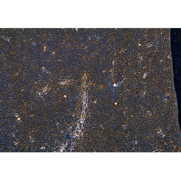
Phospho-IRF5 (Ser437) Antibody at 1/100 staining Rat thymus tissue by IHC-P. The sample was formaldehyde fixed and a heat mediated antigen retrieval step in citrate buffer was performed. The sample was then blocked and incubated with the primary antibody at 4°C overnight. An HRP conjugated anti-Rabbit antibody was used as the secondary antibody.ICC/IF
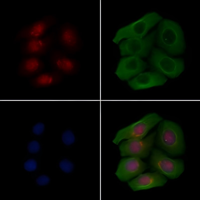
Phospho-IRF5 (Ser437) Antibody staining Hela cells(4h of LPS treatment) by IF/ICC. The samples were fixed with PFA and permeabilized in 0.1% Triton X-100,then blocked in 10% serum for 45 minutes at 25°C. Samples were then incubated with primary Ab(1:200) and mouse anti-beta tubulin Ab(1:200) for 1 hour at 37°C. An AlexaFluor594 conjugated goat anti-rabbit IgG(H+L) Ab(Red) and an AlexaFluor488 conjugated goat anti-mouse IgG(H+L) Ab(Green) were used as the secondary antibody. The nuclear counter stain is DAPI(blue).Elisa
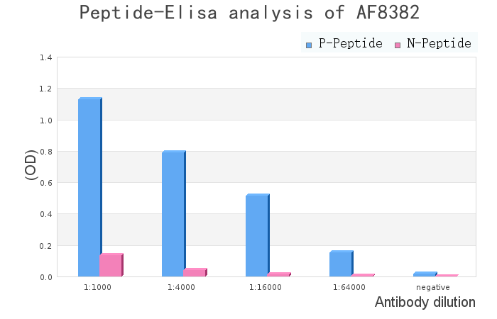
peptide-ELISA analysis of Phospho-IRF5 (Ser437) Antibody. showing specificity to antigen peptide. Peptides concentration: 1ug/ml. P-peptide: phospho-peptide. N-peptide: non-phospho-peptide.WB
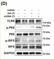
FIGURE 4 Cytoplasmic mtDNA markedly activated STING signalling. BMDCs were treated with mtDNA (10 μg/ml), or transfected with vehicle or mtDNA (10 μg/ml) for 24 h, then were harvested for western blot and qPCR. (A) Representative blots and relative quantification protein levels of STING pathway (n = 3). (B) qPCR quantitation of IFN‐β (n = 7). (C) Representative images of nuclear translocation of IRF3 in BMDC treated as indicated. Scale bar: 5 μm. (D) Representative blots and relative quantification protein levels of TLR9 pathway (n = 3). (E) qPCR quantitation of TNF‐α, IL‐6 and VCAM‐1 (n = 6–7). (F) Representative blots and relative quantification protein levels of NLRP3 pathway (n = 3). (G) qPCR quantitation of IL‐1β, IL‐18 (n = 7). Data are shown as the mean ± SEM. *p < 0.05; **p < 0.01; ***p < 0.001. BMDC, bone marrow derived dendritic cell; IFN, interferon; mtDNA, mitochondrial DNA; mtDNA (T), mtDNA transfection; Veh (T), vehicle transfection.| Product Name | Phospho-IRF5 (Ser437) Antibody |
|---|---|
| Antibody Type | Primary Antibodies |
| Immunogen | A synthesized peptide derived from human IRF5 around the phosphorylation site of Ser437. |
| Modification | p-Ser437 |
| Clonality | Polyclonal |
|---|---|
| Isotype | IgG |
| Host Species | Rabbit |
| Tested Applications | ICC/IFIHCWB |
| WB:1:1000-1:3000 IHC:1:50-1:200 ICC/IF:1:100-1:500 |
|
| Species Reactivity | HumanMouseRat |
| Concentration | 1mg/ml |
| Purification | Affinity purified |
| Gene Symbol | IRF5 |
|---|---|
| Gene Synonyms | SLEB10 |
| Gene Full Name | interferon regulatory factor 5 |
| Gene Summary | This gene encodes a member of the interferon regulatory factor (IRF) family, a group of transcription factors with diverse roles, including virus-mediated activation of interferon, and modulation of cell growth, differentiation, apoptosis, and immune system activity. Members of the IRF family are characterized by a conserved N-terminal DNA-binding domain containing tryptophan (W) repeats. Alternative promoter use and alternative splicing result in multiple transcript variants, and a 30-nt indel polymorphism (SNP rs60344245) can result in loss of a 10-aa segment. [provided by RefSeq, Dec 2016] |
| Alternative Names | Interferon regulatory factor 5 Interferon regulatory factor 5 bone marrow variant IRF 5 IRF-5 Irf5 IRF5_HUMAN SLEB10 |
| Molecular Weight(MW) | 56kDa; 56kD(Calculated). |
| Function | Transcription factor involved in the induction of interferons IFNA and INFB and inflammatory cytokines upon virus infection. Activated by TLR7 or TLR8 signaling. |
| Cellular Localization | Cytoplasm. Nucleus. |
IHC
Phospho-IRF5 (Ser437) Antibody at 1/100 staining Rat thymus tissue by IHC-P. The sample was formaldehyde fixed and a heat mediated antigen retrieval step in citrate buffer was performed. The sample was then blocked and incubated with the primary antibody at 4°C overnight. An HRP conjugated anti-Rabbit antibody was used as the secondary antibody.
ICC/IF
Phospho-IRF5 (Ser437) Antibody staining Hela cells(4h of LPS treatment) by IF/ICC. The samples were fixed with PFA and permeabilized in 0.1% Triton X-100,then blocked in 10% serum for 45 minutes at 25°C. Samples were then incubated with primary Ab(1:200) and mouse anti-beta tubulin Ab(1:200) for 1 hour at 37°C. An AlexaFluor594 conjugated goat anti-rabbit IgG(H+L) Ab(Red) and an AlexaFluor488 conjugated goat anti-mouse IgG(H+L) Ab(Green) were used as the secondary antibody. The nuclear counter stain is DAPI(blue).
Elisa
peptide-ELISA analysis of Phospho-IRF5 (Ser437) Antibody. showing specificity to antigen peptide. Peptides concentration: 1ug/ml. P-peptide: phospho-peptide. N-peptide: non-phospho-peptide.
WB
FIGURE 4 Cytoplasmic mtDNA markedly activated STING signalling. BMDCs were treated with mtDNA (10 μg/ml), or transfected with vehicle or mtDNA (10 μg/ml) for 24 h, then were harvested for western blot and qPCR. (A) Representative blots and relative quantification protein levels of STING pathway (n = 3). (B) qPCR quantitation of IFN‐β (n = 7). (C) Representative images of nuclear translocation of IRF3 in BMDC treated as indicated. Scale bar: 5 μm. (D) Representative blots and relative quantification protein levels of TLR9 pathway (n = 3). (E) qPCR quantitation of TNF‐α, IL‐6 and VCAM‐1 (n = 6–7). (F) Representative blots and relative quantification protein levels of NLRP3 pathway (n = 3). (G) qPCR quantitation of IL‐1β, IL‐18 (n = 7). Data are shown as the mean ± SEM. *p < 0.05; **p < 0.01; ***p < 0.001. BMDC, bone marrow derived dendritic cell; IFN, interferon; mtDNA, mitochondrial DNA; mtDNA (T), mtDNA transfection; Veh (T), vehicle transfection.| Application Notes | WB:1:1000-1:3000 IHC:1:50-1:200 ICC/IF:1:100-1:500 |
|---|
| Form | Liquid |
|---|---|
| Storage Instructions | Store at -20 °C. Stable for 12 months from date of receipt. |
| Storage Buffer | Rabbit IgG in phosphate buffered saline , pH 7.4, 150mM NaCl, 0.02% sodium azide and 50% glycerol. |


