WB
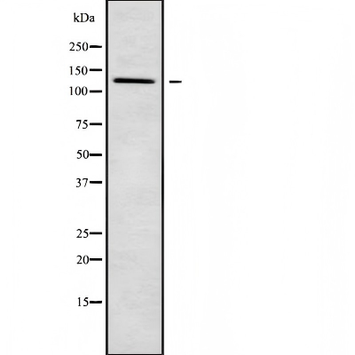
Western blot analysis of PDE3B using HuvEc whole cell lysatesIHC
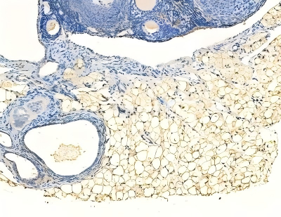
PDE3B Antibody at 1/100 staining rat muscle tissue by IHC-P. The sample was formaldehyde fixed and a heat mediated antigen retrieval step in citrate buffer was performed. The sample was then blocked and incubated with the primary antibody at 4°C overnight. An HRP conjugated anti-rabbit antibody was used as the secondary antibody.ICC/IF
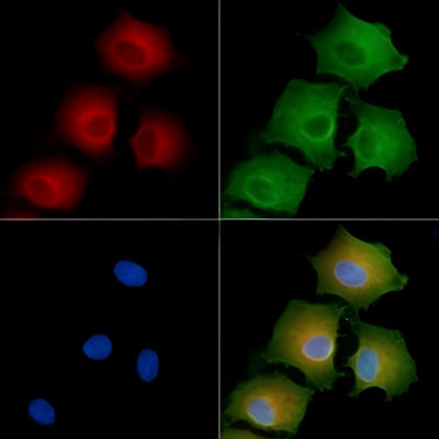
PDE3B Antibody staining A549 cells by IF/ICC. The samples were fixed with PFA and permeabilized in 0.1% Triton X-100, then blocked in 10% serum for 45 minutes at 25°C. Samples were then incubated with primary Ab and mouse anti-beta tubulin Ab for 1 hour at 37°C. An AlexaFluor594 conjugated goat anti-rabbit IgG Ab(Red) and an AlexaFluor488 conjugated goat anti-mouse IgG Ab(Green) were used as the secondary antibody. The nuclear counter stain is DAPI (blue).| Product Name | ASCL1 Antibody |
|---|---|
| Antibody Type | Primary Antibodies |
| Immunogen | A synthesized peptide derived from human ASCL1, corresponding to a region within C-terminal amino acids. |
| Clonality | Polyclonal |
|---|---|
| Isotype | IgG |
| Host Species | Rabbit |
| Tested Applications | ICC/IFIHCWB |
| WB:1:1000-1:3000 IHC:1:50-1:200 ICC/IF:1:100-1:500 |
|
| Species Reactivity | HumanMouseRat |
| Concentration | 1mg/ml |
| Purification | Affinity purified |
| Gene Symbol | ASCL1 |
|---|---|
| Gene Synonyms | ASH1 HASH1 MASH1 bHLHa46 |
| Gene Full Name | achaete-scute family bHLH transcription factor 1 |
| Gene Summary | This gene encodes a member of the basic helix-loop-helix (BHLH) family of transcription factors. The protein activates transcription by binding to the E box (5'-CANNTG-3'). Dimerization with other BHLH proteins is required for efficient DNA binding. This protein plays a role in the neuronal commitment and differentiation and in the generation of olfactory and autonomic neurons. Mutations in this gene may contribute to the congenital central hypoventilation syndrome (CCHS) phenotype in rare cases. [provided by RefSeq, Jul 2008] |
| Alternative Names | Achaete scute complex homolog 1 Achaete scute complex homolog like 1 Achaete scute complex homologue 1 Achaete scute complex homologue like 1 Achaete scute complex like 1 Achaete scute family bHLH transcription factor 1 Achaete scute protein Achaete-scute homolog 1 Ascl 1 ascl1 ASCL1_HUMAN Ash 1 ASH-1 Ash1 bHLHa46 Class A basic helix-loop-helix protein 46 Hash 1 HASH1 Mammalian achaete scute homolog 1 Mammalian achaete scute homologue 1 Mash 1 Mash1 |
| Molecular Weight(MW) | 25~35 kDa; 25kD(Calculated). |
| Cellular Localization | Membrane>Multi-pass membrane protein. |
WB
Western blot analysis of PDE3B using HuvEc whole cell lysates
IHC
PDE3B Antibody at 1/100 staining rat muscle tissue by IHC-P. The sample was formaldehyde fixed and a heat mediated antigen retrieval step in citrate buffer was performed. The sample was then blocked and incubated with the primary antibody at 4°C overnight. An HRP conjugated anti-rabbit antibody was used as the secondary antibody.
ICC/IF
PDE3B Antibody staining A549 cells by IF/ICC. The samples were fixed with PFA and permeabilized in 0.1% Triton X-100, then blocked in 10% serum for 45 minutes at 25°C. Samples were then incubated with primary Ab and mouse anti-beta tubulin Ab for 1 hour at 37°C. An AlexaFluor594 conjugated goat anti-rabbit IgG Ab(Red) and an AlexaFluor488 conjugated goat anti-mouse IgG Ab(Green) were used as the secondary antibody. The nuclear counter stain is DAPI (blue).| Application Notes | WB:1:1000-1:3000 IHC:1:50-1:200 ICC/IF:1:100-1:500 |
|---|
| Form | Liquid |
|---|---|
| Storage Instructions | Store at -20 °C. Stable for 12 months from date of receipt. |
| Storage Buffer | Rabbit IgG in phosphate buffered saline , pH 7.4, 150mM NaCl, 0.02% sodium azide and 50% glycerol. |


