WB
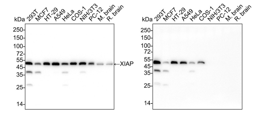
Western blot analysis of XIAP on different lysates with Rabbit anti-XIAP antibody at 1/2,000 dilution and competitor's antibody at 1/2,000 dilution. Lane 1: 293T cell lysate (20 µg/Lane), Lane 2: MCF7 cell lysate (20 µg/Lane), Lane 3: HT-29 cell lysate (20 µg/Lane), Lane 4: A549 cell lysate (20 µg/Lane), Lane 5: HeLa cell lysate (20 µg/Lane), Lane 6: COS-1 cell lysate (20 µg/Lane), Lane 7: NIH/3T3 cell lysate (20 µg/Lane), Lane 8: PC-12 cell lysate (20 µg/Lane), Lane 9: Mouse brain tissue lysate (20 µg/Lane), Lane 10: Rat brain tissue lysate (20 µg/Lane), Exposure time: Lane 1-10 (left): 20 seconds; Lane 1-10 (right): 3 minutes; ECL: K1801; 4-20% SDS-PAGE gel. Proteins were transferred to a PVDF membrane and blocked with 5% NFDM/TBST for 1 hour at room temperature. The primary antibody at 1/2,000 dilution and competitor's antibody at 1/2,000 dilution were used in 5% NFDM/TBST at 4℃ overnight. Goat Anti-Rabbit IgG - HRP Secondary Antibody at 1/50,000 dilution was used for 1 hour at room temperature.WB
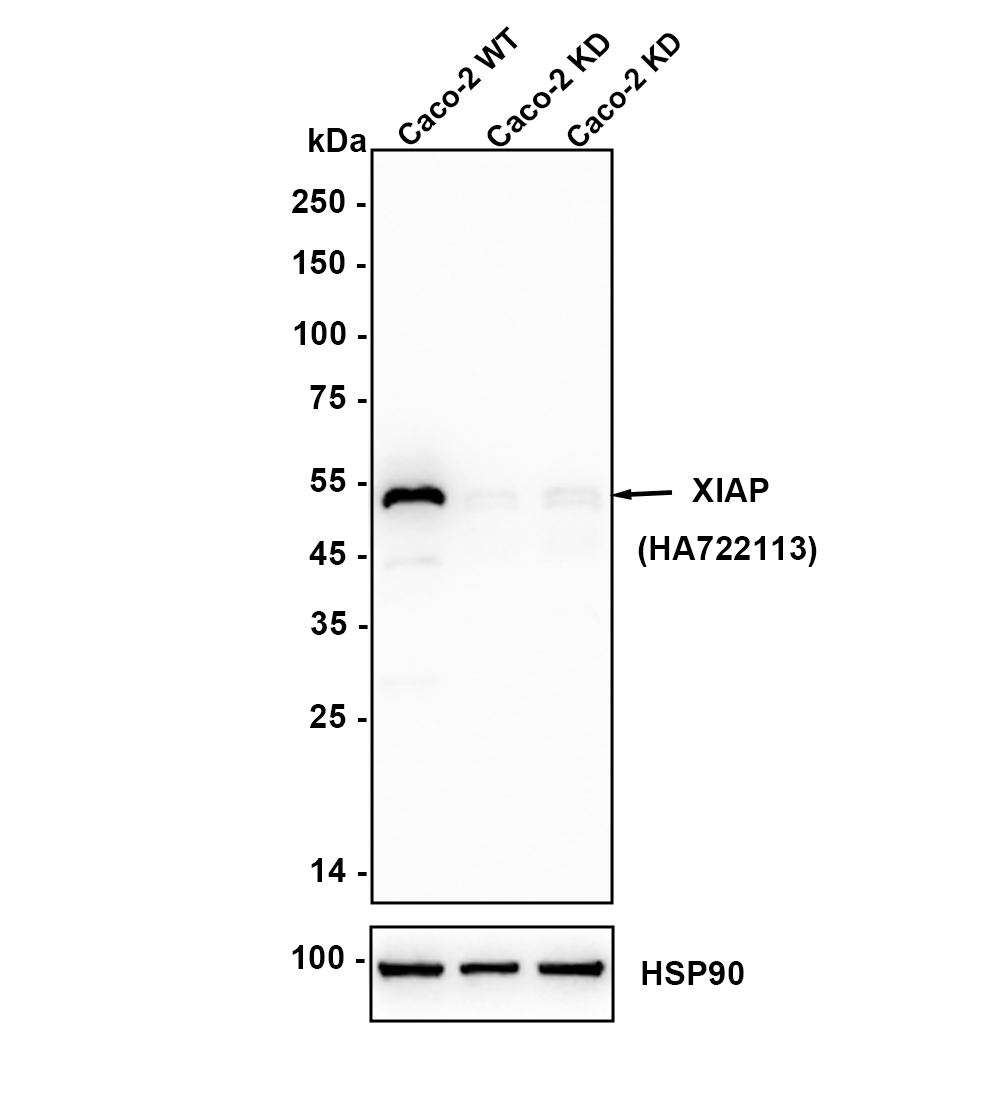
Western blot analysis of XIAP with Rabbit anti-XIAP antibody at 1/2,000 dilution. Lane 1: Wild-type Caco-2 whole cell lysate (10 µg/Lane), Lane 2/3: XIAP knockdown Caco-2 whole cell lysate (10 µg/Lane), Exposure time: 1 minute; ECL: K1801; 4-20% SDS-PAGE gel. XIAP Recombinant Rabbit Monoclonal Antibody was shown to specifically react with XIAP in wild-type Caco-2 cells. Weakened bands were observed when XIAP knockdown samples were tested. Wild-type and XIAP knockdown samples were subjected to SDS-PAGE. Proteins were transferred to a PVDF membrane and blocked with 5% NFDM in TBST for 1 hour at room temperature. The primary antibody at 1/2,000 dilution were used in 5% NFDM/TBST at 4℃ overnight. Goat Anti-Rabbit IgG - HRP Secondary Antibody at 1/50,000 dilution was used for 1 hour at room temperature.IHC
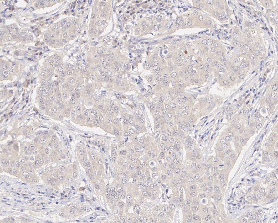
Immunohistochemical analysis of paraffin-embedded human breast cancer tissue with Rabbit anti-XIAP antibody at 1/50 dilution. The section was pre-treated using heat mediated antigen retrieval with sodium citrate buffer (pH 6.0) (high pressure) for 2 minutes. The tissues were blocked in 1% BSA for 20 minutes at room temperature, washed with ddH2O and PBS, and then probed with the primary antibody at 1/50 dilution for 1 hour at room temperature. The detection was performed using an HRP conjugated compact polymer system. DAB was used as the chromogen. Tissues were counterstained with hematoxylin and mounted with DPX.IHC
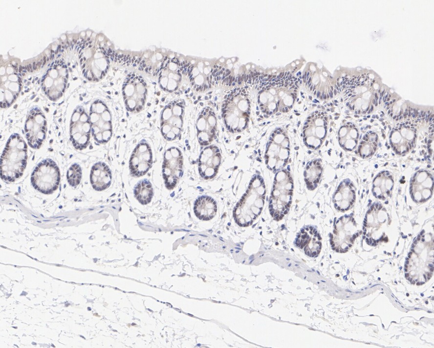
Immunohistochemical analysis of paraffin-embedded rat colon tissue with Rabbit anti-XIAP antibody at 1/50 dilution. The section was pre-treated using heat mediated antigen retrieval with sodium citrate buffer (pH 6.0) (high pressure) for 2 minutes. The tissues were blocked in 1% BSA for 20 minutes at room temperature, washed with ddH2O and PBS, and then probed with the primary antibody at 1/50 dilution for 1 hour at room temperature. The detection was performed using an HRP conjugated compact polymer system. DAB was used as the chromogen. Tissues were counterstained with hematoxylin and mounted with DPX.| Product Name | XIAP Recombinant Rabbit Monoclonal Antibody |
|---|---|
| Antibody Type | Primary Antibodies |
| Immunogen | Recombinant protein within human XIAP aa 301-450 / 497. |
| Clonality | monoclonal |
|---|---|
| Isotype | IgG |
| Host Species | Rabbit |
| Tested Applications | IHCWB |
| WB:1:2000 IHC:1:50 |
|
| Species Reactivity | HumanMonkeyMouseRat |
| Concentration | 1mg/ml |
| Purification | Protein A |
| Gene Symbol | XIAP |
|---|---|
| Gene Synonyms | API3 ILP1 MIHA XLP2 BIRC4 IAP-3 hIAP3 hIAP-3 |
| Gene Full Name | X-linked inhibitor of apoptosis |
| Gene Summary | This gene encodes a protein that belongs to a family of apoptotic suppressor proteins. Members of this family share a conserved motif termed, baculovirus IAP repeat, which is necessary for their anti-apoptotic function. This protein functions through binding to tumor necrosis factor receptor-associated factors TRAF1 and TRAF2 and inhibits apoptosis induced by menadione, a potent inducer of free radicals, and interleukin 1-beta converting enzyme. This protein also inhibits at least two members of the caspase family of cell-death proteases, caspase-3 and caspase-7. Mutations in this gene are the cause of X-linked lymphoproliferative syndrome. Alternate splicing results in multiple transcript variants. Pseudogenes of this gene are found on chromosomes 2 and 11.[provided by RefSeq, Feb 2011] |
| Alternative Names | AP 13 antibody API3 antibody Apoptosis Inhibitor 3 antibody Baculoviral IAP repeat containing 4 antibody Baculoviral IAP Repeat Containing Protein 4 antibody Baculoviral IAP repeat-containing protein 4 antibody BIRC 4 antibody BIRC4 antibody E3 ubiquitin-protein ligase XIAP antibody hIAP-3 antibody AP 13 antibody API3 antibody Apoptosis Inhibitor 3 antibody Baculoviral IAP repeat containing 4 antibody Baculoviral IAP Repeat Containing Protein 4 antibody Baculoviral IAP repeat-containing protein 4 antibody BIRC 4 antibody BIRC4 antibody E3 ubiquitin-protein ligase XIAP antibody hIAP-3 antibody hIAP3 antibody HILP antibody IAP 3 antibody IAP like protein antibody IAP-3 antibody IAP-like protein antibody IAP3 antibody ILP 1 antibody ILP antibody ILP1 antibody Inhibitor of apoptosis protein 3 antibody Inhibitor of Apoptosis X Linked antibody Mammalian IAP Homologue A antibody MIHA antibody X linked IAP antibody X linked inhibitor of apoptosis antibody X linked inhibitor of apoptosis E3 ubiquitin protein ligase antibody X linked inhibitor of apoptosis protein antibody X-linked IAP antibody x-linked inhibitor of apopotosis proteins antibody X-linked inhibitor of apoptosis protein antibody Xiap antibody XIAP_HUMAN antibody XLP2 antibody |
| Molecular Weight(MW) | 57kDa |
| Cellular Localization | Cytoplasm, Nucleus. |
WB
Western blot analysis of XIAP on different lysates with Rabbit anti-XIAP antibody at 1/2,000 dilution and competitor's antibody at 1/2,000 dilution. Lane 1: 293T cell lysate (20 µg/Lane), Lane 2: MCF7 cell lysate (20 µg/Lane), Lane 3: HT-29 cell lysate (20 µg/Lane), Lane 4: A549 cell lysate (20 µg/Lane), Lane 5: HeLa cell lysate (20 µg/Lane), Lane 6: COS-1 cell lysate (20 µg/Lane), Lane 7: NIH/3T3 cell lysate (20 µg/Lane), Lane 8: PC-12 cell lysate (20 µg/Lane), Lane 9: Mouse brain tissue lysate (20 µg/Lane), Lane 10: Rat brain tissue lysate (20 µg/Lane), Exposure time: Lane 1-10 (left): 20 seconds; Lane 1-10 (right): 3 minutes; ECL: K1801; 4-20% SDS-PAGE gel. Proteins were transferred to a PVDF membrane and blocked with 5% NFDM/TBST for 1 hour at room temperature. The primary antibody at 1/2,000 dilution and competitor's antibody at 1/2,000 dilution were used in 5% NFDM/TBST at 4℃ overnight. Goat Anti-Rabbit IgG - HRP Secondary Antibody at 1/50,000 dilution was used for 1 hour at room temperature.
WB
Western blot analysis of XIAP with Rabbit anti-XIAP antibody at 1/2,000 dilution. Lane 1: Wild-type Caco-2 whole cell lysate (10 µg/Lane), Lane 2/3: XIAP knockdown Caco-2 whole cell lysate (10 µg/Lane), Exposure time: 1 minute; ECL: K1801; 4-20% SDS-PAGE gel. XIAP Recombinant Rabbit Monoclonal Antibody was shown to specifically react with XIAP in wild-type Caco-2 cells. Weakened bands were observed when XIAP knockdown samples were tested. Wild-type and XIAP knockdown samples were subjected to SDS-PAGE. Proteins were transferred to a PVDF membrane and blocked with 5% NFDM in TBST for 1 hour at room temperature. The primary antibody at 1/2,000 dilution were used in 5% NFDM/TBST at 4℃ overnight. Goat Anti-Rabbit IgG - HRP Secondary Antibody at 1/50,000 dilution was used for 1 hour at room temperature.
IHC
Immunohistochemical analysis of paraffin-embedded human breast cancer tissue with Rabbit anti-XIAP antibody at 1/50 dilution. The section was pre-treated using heat mediated antigen retrieval with sodium citrate buffer (pH 6.0) (high pressure) for 2 minutes. The tissues were blocked in 1% BSA for 20 minutes at room temperature, washed with ddH2O and PBS, and then probed with the primary antibody at 1/50 dilution for 1 hour at room temperature. The detection was performed using an HRP conjugated compact polymer system. DAB was used as the chromogen. Tissues were counterstained with hematoxylin and mounted with DPX.
IHC
Immunohistochemical analysis of paraffin-embedded rat colon tissue with Rabbit anti-XIAP antibody at 1/50 dilution. The section was pre-treated using heat mediated antigen retrieval with sodium citrate buffer (pH 6.0) (high pressure) for 2 minutes. The tissues were blocked in 1% BSA for 20 minutes at room temperature, washed with ddH2O and PBS, and then probed with the primary antibody at 1/50 dilution for 1 hour at room temperature. The detection was performed using an HRP conjugated compact polymer system. DAB was used as the chromogen. Tissues were counterstained with hematoxylin and mounted with DPX.| Application Notes | WB:1:2000 IHC:1:50 |
|---|
| Form | Liquid |
|---|---|
| Storage Instructions | Shipped at 4℃. Store at +4℃ short term (1-2 weeks). It is recommended to aliquot into single-use upon delivery. Store at -20℃ long term. |
| Storage Buffer | PBS (pH7.4), 0.1% BSA, 40% Glycerol. Preservative: 0.05% Sodium Azide. |


