WB
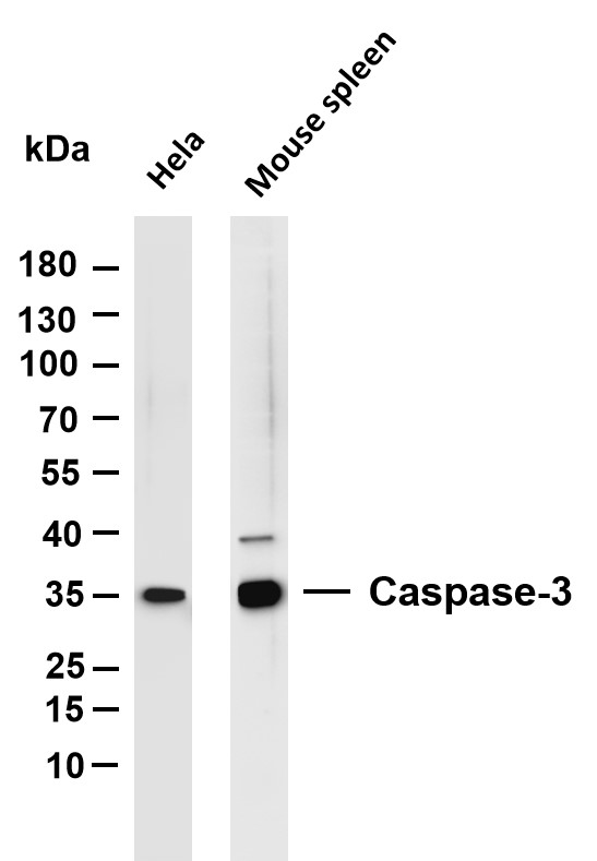
Various whole cell lysates were separated by 4-20% SDS-PAGE, and the membrane was blotted with anti-Caspase-3 antibody. The HRP-conjugated Goat anti-Rabbit IgG (H + L) antibody was used to detect the antibody. Lane 1: Hela, Lane 2: Mouse spleen,IHC
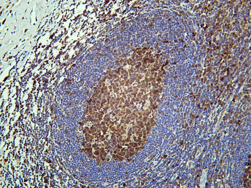
Human tonsil was stained with Anti-Caspase-3 rabbit antibodyIHC
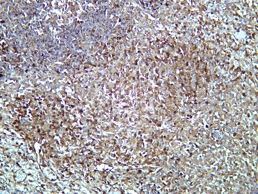
Rat spleen was stained with Anti-Caspase-3 rabbit antibodyIHC
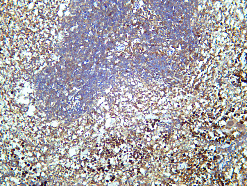
Mouse spleen was stained with Anti-Caspase-3 rabbit antibodyICC/IF
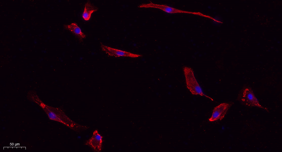
Immunofluorescence analysis of A549. 1,primary Antibody (red) was diluted at 1:200 (4℃ overnight). 2, Goat Anti Rabbit IgG (H&L) - Alexa Fluor 594 Secondary antibody was diluted at 1:1000 (room temperature, 50min).3, DAPI (blue) 10min.| Product Name | Caspase-3 Rabbit mAb |
|---|---|
| Antibody Type | Primary Antibodies |
| Immunogen | The specific immunogen used to produce this antibody is proprietary information. |
| Clonality | Monoclonal |
|---|---|
| Isotype | IgG,K |
| Host Species | Rabbit |
| Tested Applications | ICC/IFIHCWB |
| WB:1:1000-5000 IHC:1:200-1000 ICC/IF:1:200-1000 |
|
| Species Reactivity | FishHumanMouseRat |
| Concentration | 1mg/ml |
| Purification | Protein A |
| Gene Symbol | CASP3 |
|---|---|
| Gene Synonyms | CPP32 SCA-1 CPP32B |
| Gene Full Name | caspase 3 |
| Gene Summary | The protein encoded by this gene is a cysteine-aspartic acid protease that plays a central role in the execution-phase of cell apoptosis. The encoded protein cleaves and inactivates poly(ADP-ribose) polymerase while it cleaves and activates sterol regulatory element binding proteins as well as caspases 6, 7, and 9. This protein itself is processed by caspases 8, 9, and 10. It is the predominant caspase involved in the cleavage of amyloid-beta 4A precursor protein, which is associated with neuronal death in Alzheimer's disease. [provided by RefSeq, Aug 2017] |
| Alternative Names | CASP3 CPP32 Caspase-3 CASP-3 Apopain Cysteine protease CPP32 CPP-32 Protein Yama SREBP cleavage activity 1 SCA-1 |
| Molecular Weight(MW) | 35 kDa |
| Cellular Localization | Cytoplasm. |
WB
Various whole cell lysates were separated by 4-20% SDS-PAGE, and the membrane was blotted with anti-Caspase-3 antibody. The HRP-conjugated Goat anti-Rabbit IgG (H + L) antibody was used to detect the antibody. Lane 1: Hela, Lane 2: Mouse spleen,
IHC
Human tonsil was stained with Anti-Caspase-3 rabbit antibody
IHC
Rat spleen was stained with Anti-Caspase-3 rabbit antibody
IHC
Mouse spleen was stained with Anti-Caspase-3 rabbit antibody
ICC/IF
Immunofluorescence analysis of A549. 1,primary Antibody (red) was diluted at 1:200 (4℃ overnight). 2, Goat Anti Rabbit IgG (H&L) - Alexa Fluor 594 Secondary antibody was diluted at 1:1000 (room temperature, 50min).3, DAPI (blue) 10min.| Application Notes | WB:1:1000-5000 IHC:1:200-1000 ICC/IF:1:200-1000 |
|---|
| Form | Liquid |
|---|---|
| Storage Instructions | -15°C to -25°C/1 year(Do not lower than -25°C) |
| Storage Buffer | PBS, 50% glycerol, 0.05% Proclin 300, 0.05%BSA |


