WB
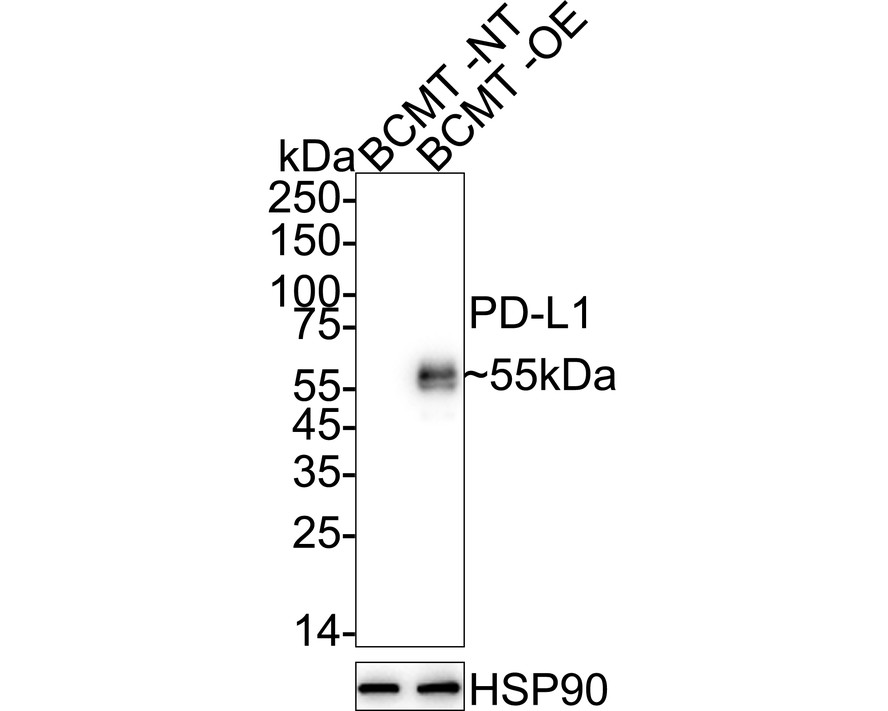
Western blot analysis of PD-L1 on different lysates with Rabbit anti-PD-L1 antibody at 1/2,000 dilution. Lane 1: BCMT cell lysate, Lane 2: BCMT transfected with PD-L1 cell lysate, Exposure time: 10 seconds; ECL: K1801; 4-20% SDS-PAGE gel. Proteins were transferred to a PVDF membrane and blocked with 5% NFDM/TBST for 1 hour at room temperature. The primary antibody at 1/2,000 dilution was used in 5% NFDM/TBST at 4℃ overnight. Goat Anti-Rabbit IgG - HRP Secondary Antibody at 1/50,000 dilution was used for 1 hour at room temperature.IHC
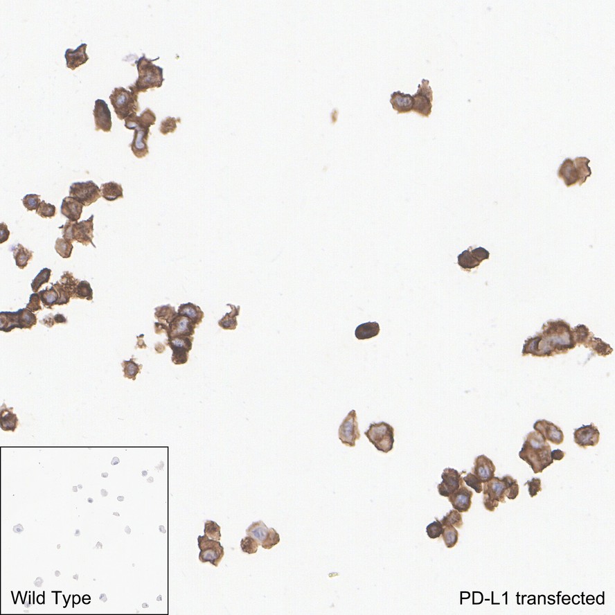
Immunohistochemical analysis of paraffin-embedded BCMT cells transfected with PD-L1 with Rabbit anti-PD-L1 antibody at 1/5,000 dilution. The section was pre-treated using heat mediated antigen retrieval with Tris-EDTA buffer (pH 9.0) for 20 minutes. The tissues were blocked in 1% BSA for 20 minutes at room temperature, washed with ddH2O and PBS, and then probed with the primary antibody at 1/5,000 dilution for 1 hour at room temperature. The detection was performed using an HRP conjugated compact polymer system. DAB was used as the chromogen. Tissues were counterstained with hematoxylin and mounted with DPX.ICC/IF
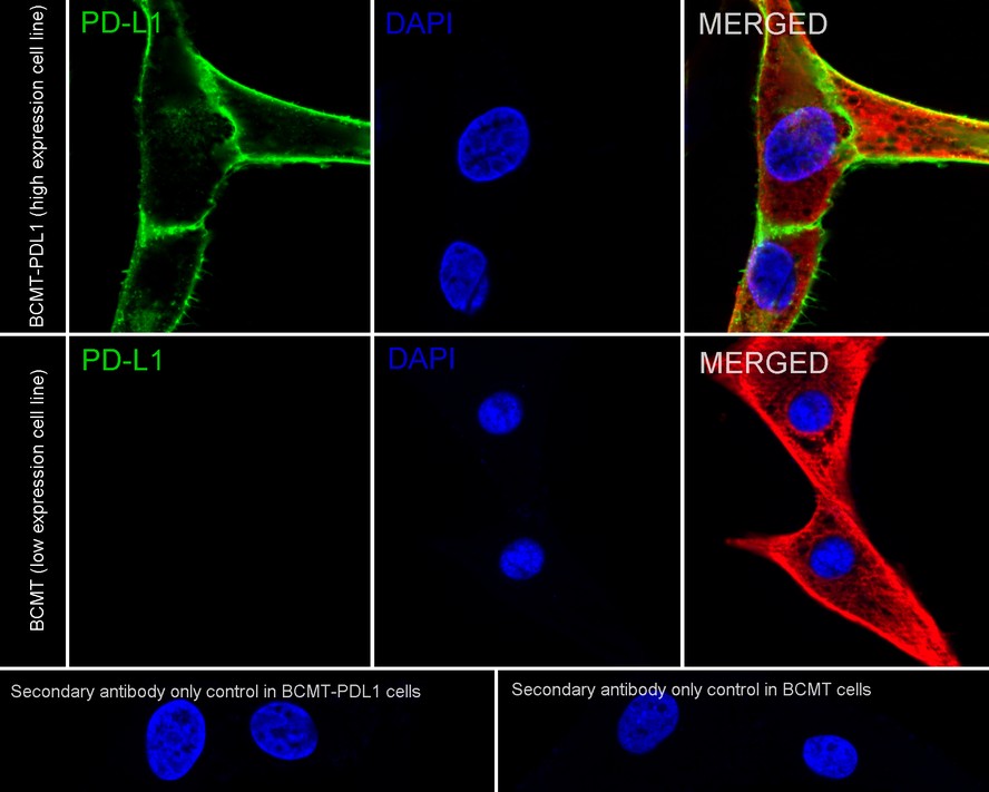
Immunocytochemistry analysis of BCMT cells transfected with PD-L1 labeling PD-L1 with Rabbit anti-PD-L1 antibody at 1/2,500 dilution. Cells were fixed in 4% paraformaldehyde for 15 minutes at room temperature, permeabilized with 0.1% Triton X-100 in PBS for 15 minutes at room temperature, then blocked with 1% BSA in 10% negative goat serum for 1 hour at room temperature. Cells were then incubated with Rabbit anti-PD-L1 antibody at 1/2,500 dilution in 1% BSA in PBST overnight at 4 ℃. Goat Anti-Rabbit IgG H&L was used as the secondary antibody at 1/1,000 dilution. PBS instead of the primary antibody was used as the secondary antibody only control. Nuclear DNA was labelled in blue with DAPI. Beta tubulin ( red) was stained at 1/100 dilution overnight at +4℃. Goat Anti-Mouse IgG H&L was used as the secondary antibody at 1/1,000 dilution.| Product Name | PD-L1 Recombinant Rabbit Monoclonal Antibody |
|---|---|
| Antibody Type | Primary Antibodies |
| Immunogen | Recombinant protein within dog PD-L1 aa 1-238. |
| Clonality | Monoclonal |
|---|---|
| Isotype | IgG |
| Host Species | Rabbit |
| Tested Applications | ICC/IFIHCWB |
| WB:1:2000 IHC:1:5000 ICC/IF:1:2500 |
|
| Species Reactivity | Dog |
| Concentration | 1mg/ml |
| Purification | Protein A |
| Gene Symbol | CD274 |
|---|---|
| Gene Synonyms | B7-H B7H1 PDL1 PD-L1 ADMIO5 hPD-L1 PDCD1L1 PDCD1LG1 |
| Gene Full Name | CD274 molecule |
| Gene Summary | This gene encodes an immune inhibitory receptor ligand that is expressed by hematopoietic and non-hematopoietic cells, such as T cells and B cells and various types of tumor cells. The encoded protein is a type I transmembrane protein that has immunoglobulin V-like and C-like domains. Interaction of this ligand with its receptor inhibits T-cell activation and cytokine production. During infection or inflammation of normal tissue, this interaction is important for preventing autoimmunity by maintaining homeostasis of the immune response. In tumor microenvironments, this interaction provides an immune escape for tumor cells through cytotoxic T-cell inactivation. Expression of this gene in tumor cells is considered to be prognostic in many types of human malignancies, including colon cancer and renal cell carcinoma. Alternative splicing results in multiple transcript variants. [provided by RefSeq, Sep 2015] |
| Target Description | Programmed death-ligand 1 (PD-L1) also known as cluster of differentiation 274 (CD274) or B7 homolog 1 (B7-H1) is a protein that in humans is encoded by the CD274 gene. Programmed death-ligand 1 (PD-L1) is a 40kDa type 1 transmembrane protein that has been speculated to play a major role in suppressing the adaptive arm of immune systems during particular events such as pregnancy, tissue allografts, autoimmune disease and other disease states such as hepatitis. Normally the adaptive immune system reacts to antigens that are associated with immune system activation by exogenous or endogenous danger signals. In turn, clonal expansion of antigen-specific CD8+ T cells and/or CD4+ helper cells is propagated. The binding of PD-L1 to the inhibitory checkpoint molecule PD-1 transmits an inhibitory signal based on interaction with phosphatases (SHP-1 or SHP-2) via Immunoreceptor Tyrosine-Based Switch Motif (ITSM). This reduces the proliferation of antigen-specific T-cells in lymph nodes, while simultaneously reducing apoptosis in regulatory T cells (anti-inflammatory, suppressive T cells) – further mediated by a lower regulation of the gene Bcl-2. PD-L1 is expressed on both hematopoietic and nonhematopoietic cells in tissues. However, the exact roles of PD-L1 on hematopoietic versus nonhematopoietic cells in modulating immune responses are unclear. |
| Alternative Names | B7 H antibody B7 H1 antibody B7 homolog 1 antibody B7-H1 antibody B7H antibody B7H1 antibody CD 274 antibody CD-274 antibody CD274 antibody CD274 antigen antibody B7 H antibody B7 H1 antibody B7 homolog 1 antibody B7-H1 antibody B7H antibody B7H1 antibody CD 274 antibody CD-274 antibody CD274 antibody CD274 antigen antibody CD274 molecule antibody MGC142294 antibody MGC142296 antibody OTTHUMP00000021029 antibody PD L1 antibody PD-L1 antibody PD1L1_HUMAN antibody PDCD1 ligand 1 antibody PDCD1L1 antibody PDCD1LG1 antibody PDL 1 antibody PDL1 antibody Programmed cell death 1 ligand 1 antibody Programmed death ligand 1 antibody RGD1566211 antibody |
| Molecular Weight(MW) | 33kDa |
| Cellular Localization | Membrane. |
WB
Western blot analysis of PD-L1 on different lysates with Rabbit anti-PD-L1 antibody at 1/2,000 dilution. Lane 1: BCMT cell lysate, Lane 2: BCMT transfected with PD-L1 cell lysate, Exposure time: 10 seconds; ECL: K1801; 4-20% SDS-PAGE gel. Proteins were transferred to a PVDF membrane and blocked with 5% NFDM/TBST for 1 hour at room temperature. The primary antibody at 1/2,000 dilution was used in 5% NFDM/TBST at 4℃ overnight. Goat Anti-Rabbit IgG - HRP Secondary Antibody at 1/50,000 dilution was used for 1 hour at room temperature.
IHC
Immunohistochemical analysis of paraffin-embedded BCMT cells transfected with PD-L1 with Rabbit anti-PD-L1 antibody at 1/5,000 dilution. The section was pre-treated using heat mediated antigen retrieval with Tris-EDTA buffer (pH 9.0) for 20 minutes. The tissues were blocked in 1% BSA for 20 minutes at room temperature, washed with ddH2O and PBS, and then probed with the primary antibody at 1/5,000 dilution for 1 hour at room temperature. The detection was performed using an HRP conjugated compact polymer system. DAB was used as the chromogen. Tissues were counterstained with hematoxylin and mounted with DPX.
ICC/IF
Immunocytochemistry analysis of BCMT cells transfected with PD-L1 labeling PD-L1 with Rabbit anti-PD-L1 antibody at 1/2,500 dilution. Cells were fixed in 4% paraformaldehyde for 15 minutes at room temperature, permeabilized with 0.1% Triton X-100 in PBS for 15 minutes at room temperature, then blocked with 1% BSA in 10% negative goat serum for 1 hour at room temperature. Cells were then incubated with Rabbit anti-PD-L1 antibody at 1/2,500 dilution in 1% BSA in PBST overnight at 4 ℃. Goat Anti-Rabbit IgG H&L was used as the secondary antibody at 1/1,000 dilution. PBS instead of the primary antibody was used as the secondary antibody only control. Nuclear DNA was labelled in blue with DAPI. Beta tubulin ( red) was stained at 1/100 dilution overnight at +4℃. Goat Anti-Mouse IgG H&L was used as the secondary antibody at 1/1,000 dilution.| Application Notes | WB:1:2000 IHC:1:5000 ICC/IF:1:2500 |
|---|
| Form | Liquid |
|---|---|
| Storage Instructions | Store at +4℃ after thawing. Aliquot store at -20℃ or -80℃. Avoid repeated freeze / thaw cycles. |
| Storage Buffer | 1*TBS (pH7.4), 0.05% BSA, 40% Glycerol. Preservative: 0.05% Sodium Azide. |


