WB
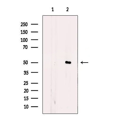
Western blot analysis of extracts from mouse brain, using CEP57 Antibody. The lane on the left was treated with blocking peptide.IHC
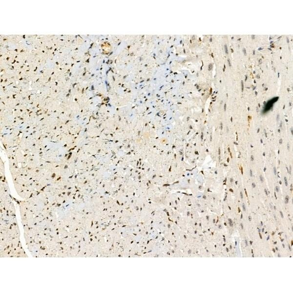
CEP57 Antibody at 1/100 staining Mouse heart tissue by IHC-P. The sample was formaldehyde fixed and a heat mediated antigen retrieval step in citrate buffer was performed. The sample was then blocked and incubated with the primary antibody at 4°C overnight. An HRP conjugated anti-Rabbit antibody was used as the secondary antibody.ICC/IF
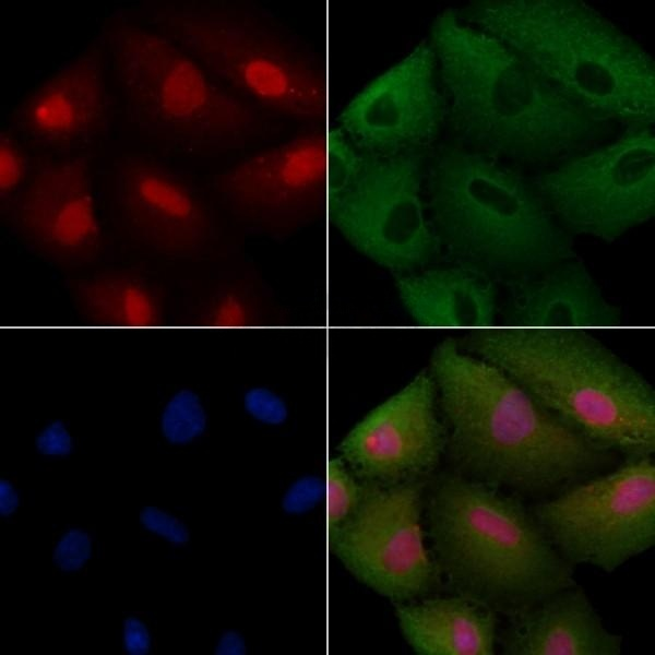
CEP57 Antibody staining A549 cells by IF/ICC. The samples were fixed with PFA and permeabilized in 0.1% Triton X-100,then blocked in 10% serum for 45 minutes at 25°C. Samples were then incubated with primary Ab and mouse anti-beta tubulin Ab for 1 hour at 37°C. An AlexaFluor594 conjugated goat anti-rabbit IgG(H+L) Ab(Red) and an AlexaFluor488 conjugated goat anti-mouse IgG(H+L) Ab(Green) were used as the secondary antibody. The nuclear counter stain is DAPI (blue).| Product Name | CEP57 Antibody |
|---|---|
| Antibody Type | Primary Antibodies |
| Immunogen | A synthesized peptide derived from human CEP57, corresponding to a region within the internal amino acids. |
| Clonality | Polyclonal |
|---|---|
| Isotype | IgG |
| Host Species | Rabbit |
| Tested Applications | ICC/IFIHCWB |
| WB:1:500-1:1000 IHC:1:50-1:200 ICC/IF:1:100-1:500 |
|
| Species Reactivity | HumanMouse |
| Concentration | 1mg/ml |
| Purification | Affinity purified |
| Gene Symbol | CEP57 |
|---|---|
| Gene Synonyms | MVA2 PIG8 TSP57 |
| Gene Full Name | centrosomal protein 57 |
| Gene Summary | This gene encodes a cytoplasmic protein called Translokin. This protein localizes to the centrosome and has a function in microtubular stabilization. The N-terminal half of this protein is required for its centrosome localization and for its multimerization, and the C-terminal half is required for nucleating, bundling and anchoring microtubules to the centrosomes. This protein specifically interacts with fibroblast growth factor 2 (FGF2), sorting nexin 6, Ran-binding protein M and the kinesins KIF3A and KIF3B, and thus mediates the nuclear translocation and mitogenic activity of the FGF2. It also interacts with cyclin D1 and controls nucleocytoplasmic distribution of the cyclin D1 in quiescent cells. This protein is crucial for maintaining correct chromosomal number during cell division. Mutations in this gene cause mosaic variegated aneuploidy syndrome, a rare autosomal recessive disorder. Multiple alternatively spliced transcript variants encoding different isoforms have been identified. [provided by RefSeq, Aug 2011] |
| Alternative Names | Centrosomal protein 57kDa Centrosomal protein of 57 kDa Cep57 Cep57 protein CEP57_HUMAN FGF2 interacting protein FGF2-interacting protein KIAA0092 MVA2 PIG8 Proliferation inducing protein 8 Testis specific protein 57 Testis-specific protein 57 Translokin TSP57 |
| Molecular Weight(MW) | 50 KD; 57kD(Calculated). |
| Cellular Localization | Nucleus. Cytoplasm. Cytoplasm>Cytoskeleton>Microtubule organizing center>Centrosome. |
WB
Western blot analysis of extracts from mouse brain, using CEP57 Antibody. The lane on the left was treated with blocking peptide.
IHC
CEP57 Antibody at 1/100 staining Mouse heart tissue by IHC-P. The sample was formaldehyde fixed and a heat mediated antigen retrieval step in citrate buffer was performed. The sample was then blocked and incubated with the primary antibody at 4°C overnight. An HRP conjugated anti-Rabbit antibody was used as the secondary antibody.
ICC/IF
CEP57 Antibody staining A549 cells by IF/ICC. The samples were fixed with PFA and permeabilized in 0.1% Triton X-100,then blocked in 10% serum for 45 minutes at 25°C. Samples were then incubated with primary Ab and mouse anti-beta tubulin Ab for 1 hour at 37°C. An AlexaFluor594 conjugated goat anti-rabbit IgG(H+L) Ab(Red) and an AlexaFluor488 conjugated goat anti-mouse IgG(H+L) Ab(Green) were used as the secondary antibody. The nuclear counter stain is DAPI (blue).| Application Notes | WB:1:500-1:1000 IHC:1:50-1:200 ICC/IF:1:100-1:500 |
|---|
| Form | Liquid |
|---|---|
| Storage Instructions | Store at -20 °C. Stable for 12 months from date of receipt. |
| Storage Buffer | Rabbit IgG in phosphate buffered saline , pH 7.4, 150mM NaCl, 0.02% sodium azide and 50% glycerol. |


