WB
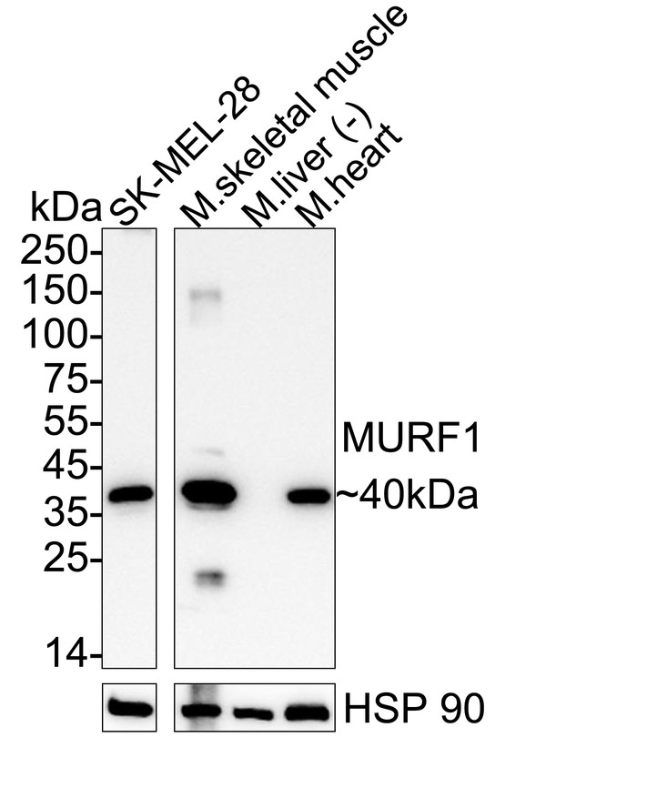
Western blot analysis of MURF1 on different lysates with Rabbit anti-MURF1 antibody at 1/5,000 dilution. Lane 1: SK-MEL-28 cell lysate (20 µg/Lane), Lane 2: Mouse skeletal muscle tissue lysate (40 µg/Lane), Lane 3: Mouse liver tissue lysate (negative) (40 µg/Lane), Lane 4: Mouse heart tissue lysate (40 µg/Lane), Exposure time:1 minute 3 seconds; ECL: K1801; 4-20% SDS-PAGE gel. Proteins were transferred to a PVDF membrane and blocked with 5% NFDM/TBST for 1 hour at room temperature. The primary antibody at 1/5,000 dilution was used in primary antibody dilution at 4℃ overnight. Goat Anti-Rabbit IgG - HRP Secondary Antibody at 1/50,000 dilution was used for 1 hour at room temperature.IP
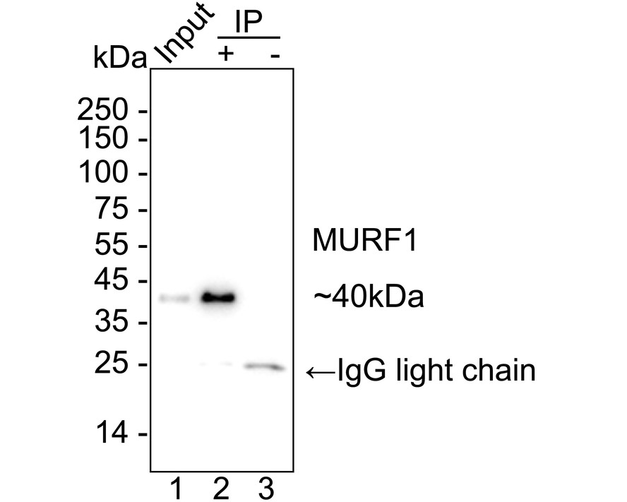
MURF1 was immunoprecipitated from 0.2 mg SK-MEL-28 cell lysate with MURF1 at 2 µg/10 µl beads. Western blot was performed from the immunoprecipitate using MURF1 at 1/5,000 dilution. HRP Conjugated Anti-Rabbit IgG for IP Nano-secondary antibody at 1/5,000 dilution was used for 1 hour at room temperature. Lane 1: SK-MEL-28 cell lysate (input) Lane 2: IP in SK-MEL-28 cell lysate Lane 3: Rabbit IgG instead of MURF1 in SK-MEL-28 cell lysate Blocking/Dilution buffer: primary antibody dilution Exposure time: 10 seconds;FC
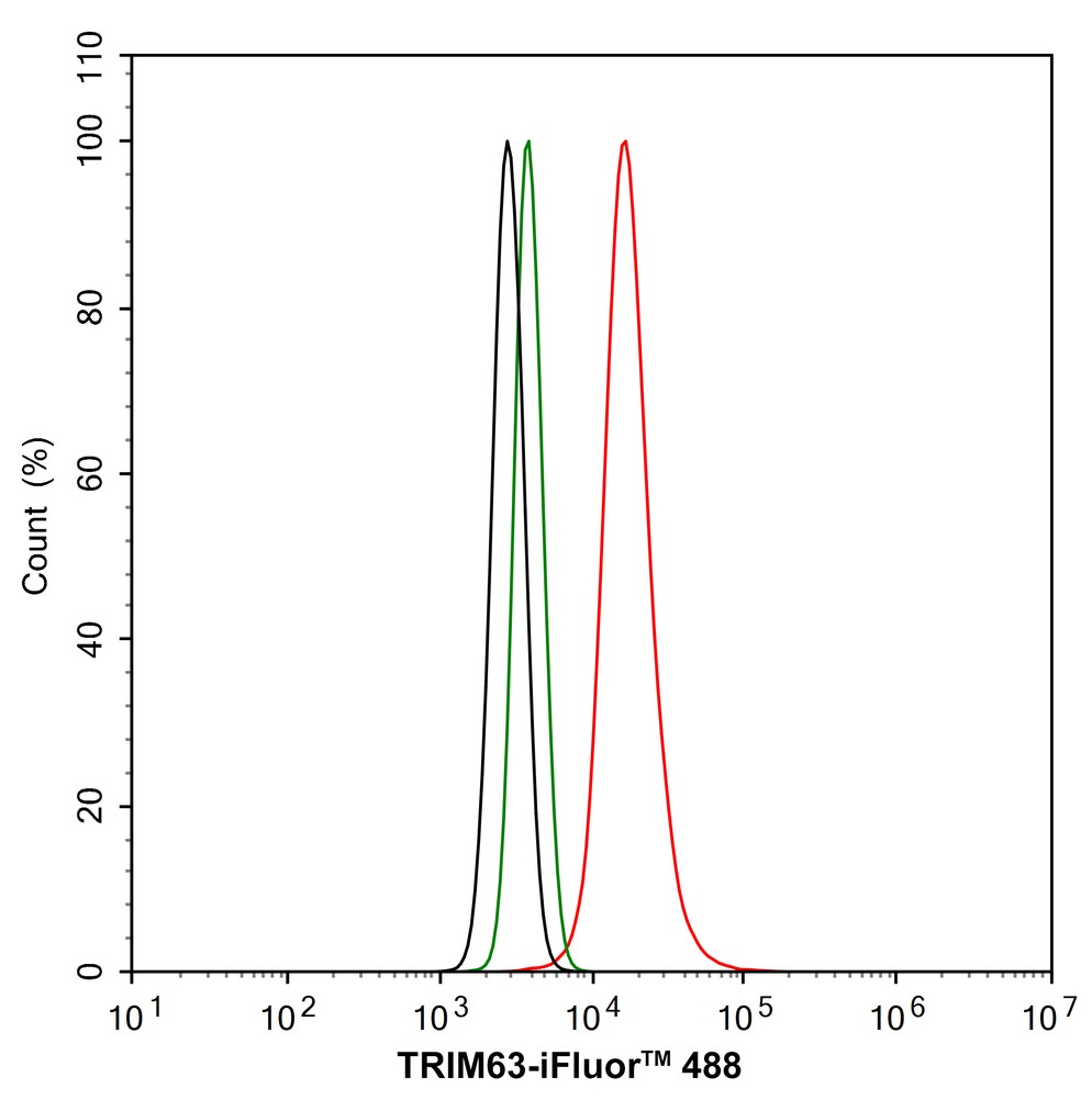
Flow cytometric analysis of SK-MEL-28 cells labeling MURF1. Cells were fixed and permeabilized. Then stained with the primary antibody (1/1,000) (red) compared with Rabbit IgG Isotype Control (green). After incubation of the primary antibody at +4℃ for an hour, the cells were stained with a iFluor™ 488 conjugate-Goat anti-Rabbit IgG Secondary antibody at 1/1,000 dilution for 30 minutes at +4℃. Unlabelled sample was used as a control (cells without incubation with primary antibody; black).| Product Name | MURF1 Recombinant Rabbit Monoclonal Antibody |
|---|---|
| Antibody Type | Primary Antibodies |
| Immunogen | Recombinant protein within human MURF1 aa 180-343 /353 |
| Clonality | Monoclonal |
|---|---|
| Isotype | IgG |
| Host Species | Rabbit |
| Tested Applications | FCIPWB |
| WB:1:5000 IP:1-2μg/sample |
|
| Species Reactivity | HumanMouseRat |
| Concentration | 1mg/ml |
| Purification | Protein A |
| Gene Symbol | TRIM63 |
|---|---|
| Gene Synonyms | IRF SMRZ CMH31 MURF1 MURF2 RNF28 |
| Gene Full Name | tripartite motif containing 63 |
| Gene Summary | This gene encodes a member of the RING zinc finger protein family found in striated muscle and iris. The product of this gene is an E3 ubiquitin ligase that localizes to the Z-line and M-line lattices of myofibrils. This protein plays an important role in the atrophy of skeletal and cardiac muscle and is required for the degradation of myosin heavy chain proteins, myosin light chain, myosin binding protein, and for muscle-type creatine kinase. [provided by RefSeq, Feb 2012] |
| Alternative Names | IRF antibody MURF1 antibody RNF28 antibody SMRZ antibody TRIM63 antibody E3 ubiquitin-protein ligase TRIM63 antibody Iris RING finger protein antibody Muscle-specific RING finger protein 1 antibody RING finger protein 28 antibody RING-type E3 ubiquitin transferase TRIM63 antibody IRF antibody MURF1 antibody RNF28 antibody SMRZ antibody TRIM63 antibody E3 ubiquitin-protein ligase TRIM63 antibody Iris RING finger protein antibody Muscle-specific RING finger protein 1 antibody RING finger protein 28 antibody RING-type E3 ubiquitin transferase TRIM63 antibody Striated muscle RING zinc finger protein antibody Tripartite motif-containing protein 63 antibody MuRF-1 antibody MuRF1 antibody |
| Molecular Weight(MW) | Predicted band size: 40 kDa |
| Cellular Localization | Cytoplasm, Nucleus, myofibril, sarcomere, M line, Z line. |
WB
Western blot analysis of MURF1 on different lysates with Rabbit anti-MURF1 antibody at 1/5,000 dilution. Lane 1: SK-MEL-28 cell lysate (20 µg/Lane), Lane 2: Mouse skeletal muscle tissue lysate (40 µg/Lane), Lane 3: Mouse liver tissue lysate (negative) (40 µg/Lane), Lane 4: Mouse heart tissue lysate (40 µg/Lane), Exposure time:1 minute 3 seconds; ECL: K1801; 4-20% SDS-PAGE gel. Proteins were transferred to a PVDF membrane and blocked with 5% NFDM/TBST for 1 hour at room temperature. The primary antibody at 1/5,000 dilution was used in primary antibody dilution at 4℃ overnight. Goat Anti-Rabbit IgG - HRP Secondary Antibody at 1/50,000 dilution was used for 1 hour at room temperature.
IP
MURF1 was immunoprecipitated from 0.2 mg SK-MEL-28 cell lysate with MURF1 at 2 µg/10 µl beads. Western blot was performed from the immunoprecipitate using MURF1 at 1/5,000 dilution. HRP Conjugated Anti-Rabbit IgG for IP Nano-secondary antibody at 1/5,000 dilution was used for 1 hour at room temperature. Lane 1: SK-MEL-28 cell lysate (input) Lane 2: IP in SK-MEL-28 cell lysate Lane 3: Rabbit IgG instead of MURF1 in SK-MEL-28 cell lysate Blocking/Dilution buffer: primary antibody dilution Exposure time: 10 seconds;
FC
Flow cytometric analysis of SK-MEL-28 cells labeling MURF1. Cells were fixed and permeabilized. Then stained with the primary antibody (1/1,000) (red) compared with Rabbit IgG Isotype Control (green). After incubation of the primary antibody at +4℃ for an hour, the cells were stained with a iFluor™ 488 conjugate-Goat anti-Rabbit IgG Secondary antibody at 1/1,000 dilution for 30 minutes at +4℃. Unlabelled sample was used as a control (cells without incubation with primary antibody; black).| Application Notes | WB:1:5000 IP:1-2μg/sample |
|---|
| Form | Liquid |
|---|---|
| Storage Instructions | Store at +4℃ after thawing. Aliquot store at -20℃ or -80℃. Avoid repeated freeze / thaw cycles. |
| Storage Buffer | 1*TBS (pH7.4), 0.05% BSA, 40% Glycerol. Preservative: 0.05% Sodium Azide. |


