WB
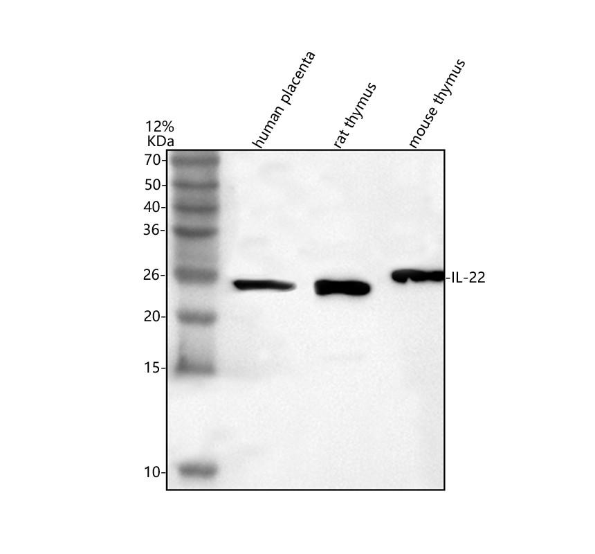
Western blot analysis of IL22 using anti-IL22 antibody. The sample well of each lane was loaded with 30 ug of sample under reducing conditions. Lane 1: human placenta tissue lysates, Lane 2: rat thymus tissue lysates, Lane 3: mouse thymus tissue lysates. After electrophoresis, proteins were transferred to a membrane. Then the membrane was incubated with rabbit anti-IL22 antigen affinity purified polyclonal antibody at a dilution of 1:1000 and probed with a goat anti-rabbit IgG-HRP secondary antibody. The signal is developed using ECL Plus Western Blotting Substrate. A specific band was detected for IL22 at approximately 24 kDa. The expected band size for IL22 is at 20 kDa.IHC
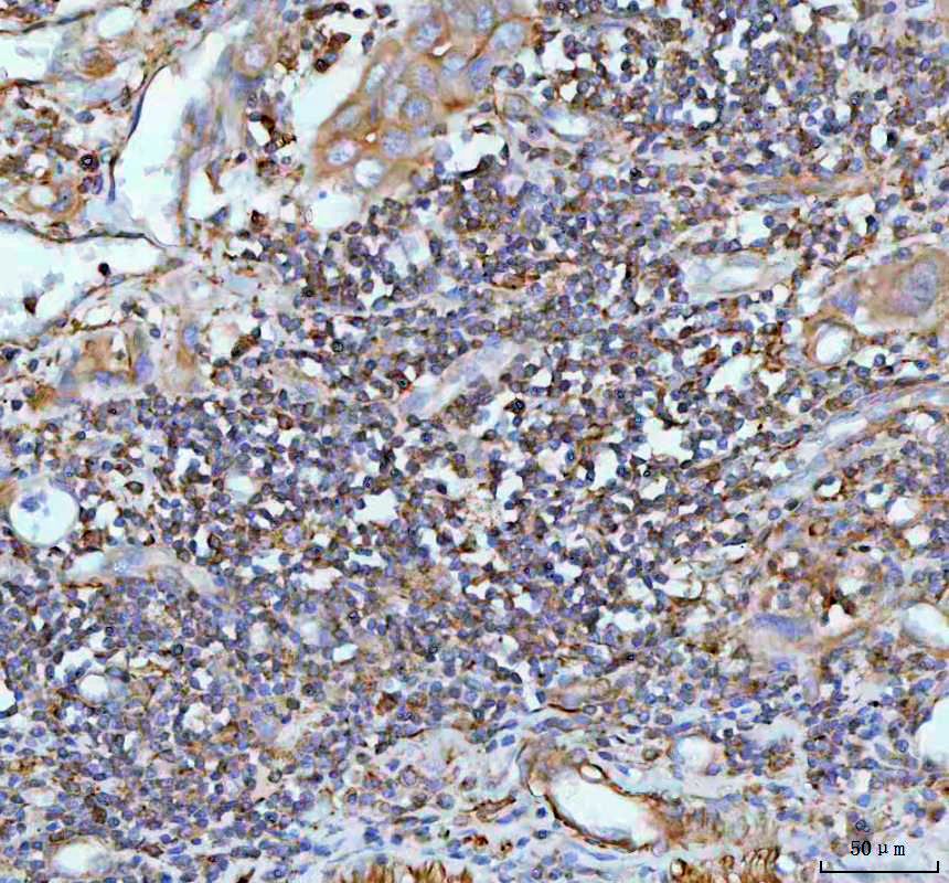
IHC analysis of IL22 using anti-IL22 antibody. IL22 was detected in a paraffin-embedded section of human rectum adenocarcinoma tissue. The tissue section was incubated with rabbit anti-IL22 Antibody at a dilution of 1:200 and developed using HRP Conjugated Rabbit IgG Super Vision Assay Kit with DAB as the chromogen.IHC
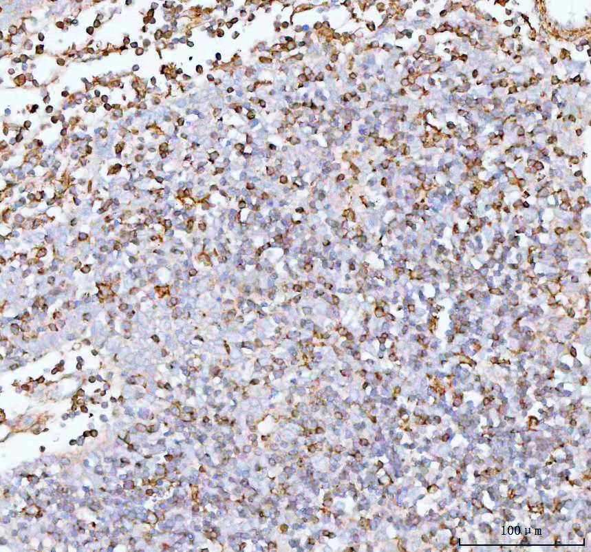
IHC analysis of IL22 using anti-IL22 antibody. IL22 was detected in a paraffin-embedded section of human spleen tissue. The tissue section was incubated with rabbit anti-IL22 Antibody at a dilution of 1:200 and developed using HRP Conjugated Rabbit IgG Super Vision Assay Kit with DAB as the chromogen.| Product Name | Anti-IL22 Antibody |
|---|---|
| Antibody Type | Primary Antibodies |
| Immunogen | E. coli-derived human IL-22 recombinant protein (Position: A34-I179). Human IL-22 shares 81.4% amino acid (aa) sequence identity with mouse IL-22. |
| Clonality | Polyclonal |
|---|---|
| Isotype | IgG |
| Host Species | Rabbit |
| Tested Applications | IHCWB |
| WB:1:500-1:2000 IHC:1:50-1:40 |
|
| Species Reactivity | HumanMouseRat |
| Concentration | 0.5mg/ml |
| Purification | Affinity purified |
| Gene Symbol | IL22 |
|---|---|
| Gene Synonyms | TIFa IL-21 IL-22 ILTIF IL-TIF IL-D110 zcyto18 TIFIL-23 |
| Gene Full Name | interleukin 22 |
| Gene Summary | This gene is a member of the IL10 family of cytokines that mediate cellular inflammatory responses. The encoded protein functions in antimicrobial defense at mucosal surfaces and in tissue repair. This protein also has pro-inflammatory properties and plays a role in in the pathogenesis of several intestinal diseases. The encoded protein is a crucial cytokine that regulates host immunity in infectious diseases, including COVID-19 (disease caused by SARS-CoV-2). [provided by RefSeq, Dec 2021] |
| Alternative Names | Cytokine Zcyto18 IL 21 IL 22 IL D110 IL TIF IL21 IL22 IL-22 ILTIF interleukin 22 TIFa TIFIL 23 zcyto18 |
| Molecular Weight(MW) | 20kDa(Observed MW 20kDa) |
| Cellular Localization | Secreted |
WB
Western blot analysis of IL22 using anti-IL22 antibody. The sample well of each lane was loaded with 30 ug of sample under reducing conditions. Lane 1: human placenta tissue lysates, Lane 2: rat thymus tissue lysates, Lane 3: mouse thymus tissue lysates. After electrophoresis, proteins were transferred to a membrane. Then the membrane was incubated with rabbit anti-IL22 antigen affinity purified polyclonal antibody at a dilution of 1:1000 and probed with a goat anti-rabbit IgG-HRP secondary antibody. The signal is developed using ECL Plus Western Blotting Substrate. A specific band was detected for IL22 at approximately 24 kDa. The expected band size for IL22 is at 20 kDa.
IHC
IHC analysis of IL22 using anti-IL22 antibody. IL22 was detected in a paraffin-embedded section of human rectum adenocarcinoma tissue. The tissue section was incubated with rabbit anti-IL22 Antibody at a dilution of 1:200 and developed using HRP Conjugated Rabbit IgG Super Vision Assay Kit with DAB as the chromogen.
IHC
IHC analysis of IL22 using anti-IL22 antibody. IL22 was detected in a paraffin-embedded section of human spleen tissue. The tissue section was incubated with rabbit anti-IL22 Antibody at a dilution of 1:200 and developed using HRP Conjugated Rabbit IgG Super Vision Assay Kit with DAB as the chromogen.| Application Notes | WB:1:500-1:2000 IHC:1:50-1:40 |
|---|
| Form | Liquid |
|---|---|
| Storage Instructions | 12 months from date of receipt,-20℃ as supplied. 6 months 2 to 8℃ after reconstitution. Avoid repeated freezing and thawing. |
| Storage Buffer | 0.5mg/ml antibody with PBS, 0.02% NaN3, 1 mg/ml BSA and 50% glycerol. |


