WB
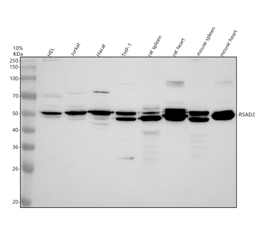
Western blot analysis of RSAD2 using anti-RSAD2 antibody . The sample well of each lane was loaded with 30 ug of sample under reducing conditions. Lane 1: human HEL whole cell lysates, Lane 2: human Jurkat whole cell lysates, Lane 3: human Hacat whole cell lysates, Lane 4: human THP-1 whole cell lysates, Lane 5: rat spleen tissue lysates, Lane 6: rat heart tissue lysates, Lane 7: mouse spleen tissue lysates, Lane 8: mouse heart tissue lysates. After electrophoresis, proteins were transferred to a membrane. Then the membrane was incubated with rabbit anti-RSAD2 antigen affinity purified polyclonal antibody at a dilution of 1:1000 and probed with a goat anti-rabbit IgG-HRP secondary antibody (Catalog # BA1054). The signal is developed using ECL Plus Western Blotting Substrate (Catalog # AR1197). A specific band was detected for RSAD2 at approximately 50 kDa. The expected band size for RSAD2 is at 42 kDa.IHC
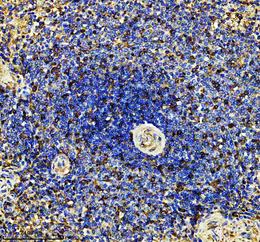
IHC analysis of RSAD2 using anti-RSAD2 antibody . RSAD2 was detected in a paraffin-embedded section of human spleen tissue. The tissue section was incubated with rabbit anti-RSAD2 Antibody at a dilution of 1:200 and developed using HRP Conjugated Rabbit IgG Super Vision Assay Kit (Catalog # SV0002) with DAB (Catalog # AR1027) as the chromogen.IHC
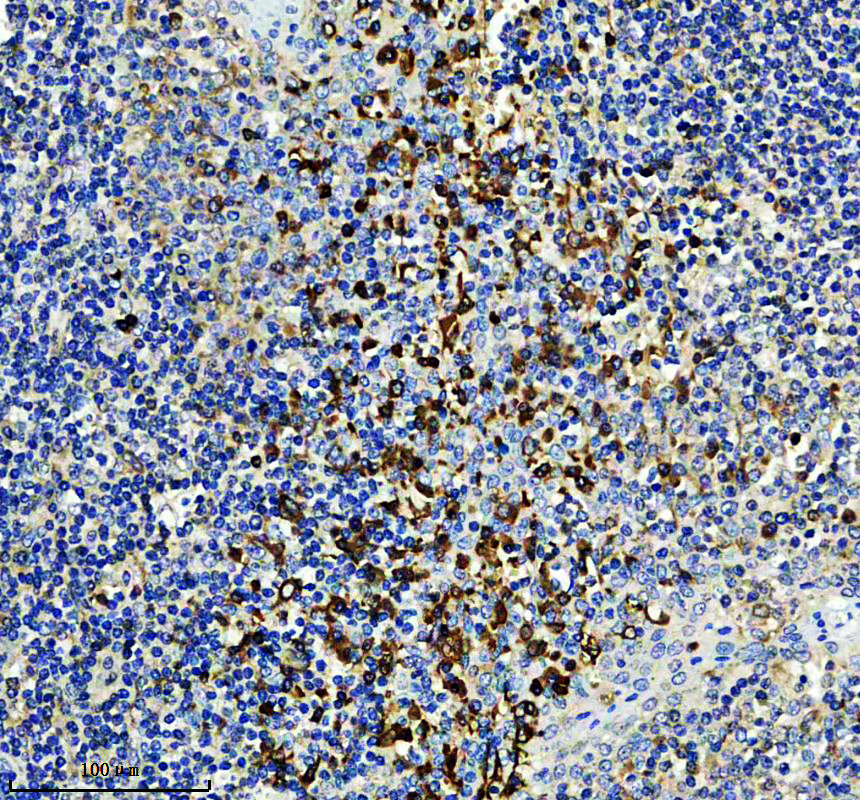
IHC analysis of RSAD2 using anti-RSAD2 antibody . RSAD2 was detected in a paraffin-embedded section of human tonsil tissue. The tissue section was incubated with rabbit anti-RSAD2 Antibody at a dilution of 1:200 and developed using HRP Conjugated Rabbit IgG Super Vision Assay Kit (Catalog # SV0002) with DAB (Catalog # AR1027) as the chromogen.ICC/IF
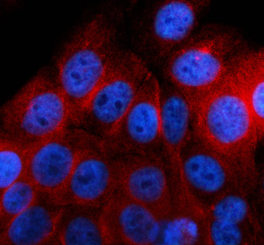
ICC/IF analysis of RSAD2 using anti-RSAD2 antibody . RSAD2 was detected in an immunocytochemical section of Hacat cells. The section was incubated with rabbit anti-RSAD2 Antibody at a dilution of 1:100. Cy3-conjugated Anti-rabbit IgG Secondary Antibody (red) (Catalog # BA1032) was used as secondary antibody. The section was counterstained with DAPI (Catalog # AR1176) (Blue).| Product Name | Anti-RSAD2 Antibody |
|---|---|
| Antibody Type | Primary Antibodies |
| Immunogen | E.coli-derived human RSAD2 recombinant protein (Position: A43-D299). |
| Clonality | Polyclonal |
|---|---|
| Isotype | IgG |
| Host Species | Rabbit |
| Tested Applications | ELISAICC/IFIHCWB |
| WB:1:500-2000 IHC:1:50-400 ICC/IF:1:50-400 ELISA:1:100-1000 |
|
| Species Reactivity | HumanMouseRat |
| Concentration | 0.5mg/ml |
| Purification | Affinity purified |
| Gene Symbol | RSAD2 |
|---|---|
| Gene Synonyms | SAND cig5 vig1 cig33 |
| Gene Full Name | radical S-adenosyl methionine domain containing 2 |
| Gene Summary | The protein encoded by this gene is an interferon-inducible antiviral protein that belongs to the S-adenosyl-L-methionine (SAM) superfamily of enzymes. The protein plays a role in cellular antiviral response and innate immune signaling. Antiviral effects result from inhibition of viral RNA replication, interference in the secretory pathway, binding to viral proteins and dysregulation of cellular lipid metabolism. The protein has been found to inhibit both DNA and RNA viruses, including influenza virus, human immunodeficiency virus (HIV-1) and Zika virus. [provided by RefSeq, Sep 2020] |
| Alternative Names | RSAD2 CIG5 Radical S-adenosyl methionine domain-containing protein 2 Cytomegalovirus-induced gene 5 protein Viperin Virus inhibitory protein endoplasmic reticulum-associated interferon-inducible |
| Molecular Weight(MW) | 50 kDa |
| Cellular Localization | Endoplasmic reticulum,Golgi apparatus,Lipid droplet,Membrane,Mitochondrion |
WB
Western blot analysis of RSAD2 using anti-RSAD2 antibody . The sample well of each lane was loaded with 30 ug of sample under reducing conditions. Lane 1: human HEL whole cell lysates, Lane 2: human Jurkat whole cell lysates, Lane 3: human Hacat whole cell lysates, Lane 4: human THP-1 whole cell lysates, Lane 5: rat spleen tissue lysates, Lane 6: rat heart tissue lysates, Lane 7: mouse spleen tissue lysates, Lane 8: mouse heart tissue lysates. After electrophoresis, proteins were transferred to a membrane. Then the membrane was incubated with rabbit anti-RSAD2 antigen affinity purified polyclonal antibody at a dilution of 1:1000 and probed with a goat anti-rabbit IgG-HRP secondary antibody (Catalog # BA1054). The signal is developed using ECL Plus Western Blotting Substrate (Catalog # AR1197). A specific band was detected for RSAD2 at approximately 50 kDa. The expected band size for RSAD2 is at 42 kDa.
IHC
IHC analysis of RSAD2 using anti-RSAD2 antibody . RSAD2 was detected in a paraffin-embedded section of human spleen tissue. The tissue section was incubated with rabbit anti-RSAD2 Antibody at a dilution of 1:200 and developed using HRP Conjugated Rabbit IgG Super Vision Assay Kit (Catalog # SV0002) with DAB (Catalog # AR1027) as the chromogen.
IHC
IHC analysis of RSAD2 using anti-RSAD2 antibody . RSAD2 was detected in a paraffin-embedded section of human tonsil tissue. The tissue section was incubated with rabbit anti-RSAD2 Antibody at a dilution of 1:200 and developed using HRP Conjugated Rabbit IgG Super Vision Assay Kit (Catalog # SV0002) with DAB (Catalog # AR1027) as the chromogen.
ICC/IF
ICC/IF analysis of RSAD2 using anti-RSAD2 antibody . RSAD2 was detected in an immunocytochemical section of Hacat cells. The section was incubated with rabbit anti-RSAD2 Antibody at a dilution of 1:100. Cy3-conjugated Anti-rabbit IgG Secondary Antibody (red) (Catalog # BA1032) was used as secondary antibody. The section was counterstained with DAPI (Catalog # AR1176) (Blue).| Application Notes | WB:1:500-2000 IHC:1:50-400 ICC/IF:1:50-400 ELISA:1:100-1000 |
|---|
| Form | Liquid |
|---|---|
| Storage Instructions | 12 months from date of receipt, -20℃ as supplied. 6 months 2 to 8℃ after reconstitution. Avoid repeated freezing and thawing. |
| Storage Buffer | 500 ug/ml antibody with PBS, 0.02% NaN3, 1 mg/ml BSA and 50% glycerol. |


