WB
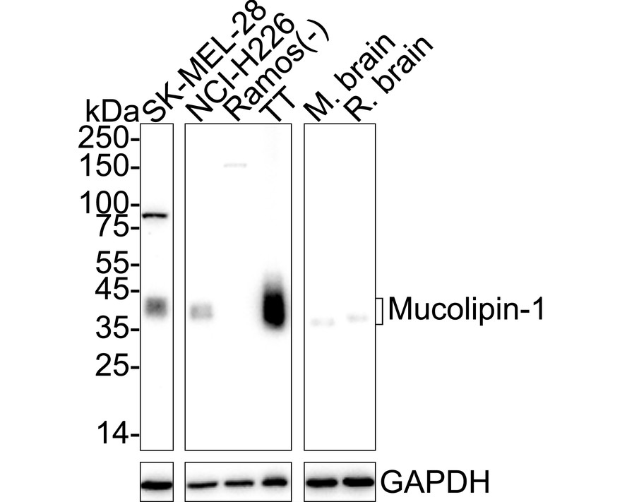
Western blot analysis of Mucolipin-1 on different lysates with Rabbit anti-Mucolipin-1 antibody at 1/2,000 dilution. Lane 1: SK-MEL-28 cell lysate Lane 2: NCI-H226 cell lysate Lane 3: Ramos cell lysate (negative) Lane 4: TT cell lysate Lane 5: Mouse brain tissue lysate Lane 6: Rat brain tissue lysate Lysates/proteins at 20 µg/Lane. Predicted band size: 65 kDa Observed band size: 35-40 kDa (PMID: 16257972) Exposure time: 3 minutes; ECL: K1801; 4-20% SDS-PAGE gel. Proteins were transferred to a PVDF membrane and blocked with 5% NFDM/TBST for 1 hour at room temperature. The primary antibody at 1/2,000 dilution was used in 5% NFDM/TBST at 4℃ overnight. Goat Anti-Rabbit IgG - HRP Secondary Antibody at 1/50,000 dilution was used for 1 hour at room temperature.IHC
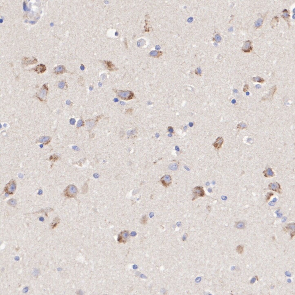
Immunohistochemical analysis of paraffin-embedded human brain tissue with Rabbit anti-Mucolipin-1 antibody at 1/1,000 dilution. The section was pre-treated using heat mediated antigen retrieval with Tris-EDTA buffer (pH 9.0) for 20 minutes. The tissues were blocked in 1% BSA for 20 minutes at room temperature, washed with ddH2O and PBS, and then probed with the primary antibody at 1/1,000 dilution for 1 hour at room temperature. The detection was performed using an HRP conjugated compact polymer system. DAB was used as the chromogen. Tissues were counterstained with hematoxylin and mounted with DPX.| Product Name | Mucolipin-1 Recombinant Rabbit Monoclonal Antibody |
|---|---|
| Antibody Type | Primary Antibodies |
| Immunogen | Recombinant protein within human Mucolipin-1 aa 66-321. |
| Clonality | Monoclonal |
|---|---|
| Isotype | IgG |
| Host Species | Rabbit |
| Tested Applications | IHCWB |
| WB:1:2,000 IHC:1:1,000 |
|
| Species Reactivity | HumanMouseRat |
| Concentration | 1mg/ml |
| Purification | Protein A |
| Gene Symbol | MCOLN1 |
|---|---|
| Gene Synonyms | ML1 ML4 LECD MG-2 MLIV MST080 TRPML1 MSTP080 TRP-ML1 TRPM-L1 |
| Gene Full Name | mucolipin TRP cation channel 1 |
| Gene Summary | This gene encodes a memberof the transient receptor potential (TRP) cation channel gene family. The transmembrane protein localizes to intracellular vesicular membranes including lysosomes, and functions in the late endocytic pathway and in the regulation of lysosomal exocytosis. The channel is permeable to Ca(2+), Fe(2+), Na(+), K(+), and H(+), and is modulated by changes in Ca(2+) concentration. Mutations in this gene result in mucolipidosis type IV. [provided by RefSeq, Oct 2009] |
| Alternative Names | MCLN1_HUMAN antibody Mcoln 1 antibody Mcoln1 antibody MG 2 antibody MG-2 antibody MG2 antibody ML 4 antibody ML4 antibody MLIV antibody MST080 antibody MCLN1_HUMAN antibody Mcoln 1 antibody Mcoln1 antibody MG 2 antibody MG-2 antibody MG2 antibody ML 4 antibody ML4 antibody MLIV antibody MST080 antibody MSTP080 antibody Mucolipidin antibody Mucolipin 1 antibody Mucolipin-1 antibody Mucolipin1 antibody TRP ML1 antibody TRPML1 antibody |
| Molecular Weight(MW) | 65 kDa |
| Cellular Localization | Cell membrane,Cell projection,Cytoplasmic vesicle,Endosome,Lysosome,Membrane |
WB
Western blot analysis of Mucolipin-1 on different lysates with Rabbit anti-Mucolipin-1 antibody at 1/2,000 dilution. Lane 1: SK-MEL-28 cell lysate Lane 2: NCI-H226 cell lysate Lane 3: Ramos cell lysate (negative) Lane 4: TT cell lysate Lane 5: Mouse brain tissue lysate Lane 6: Rat brain tissue lysate Lysates/proteins at 20 µg/Lane. Predicted band size: 65 kDa Observed band size: 35-40 kDa (PMID: 16257972) Exposure time: 3 minutes; ECL: K1801; 4-20% SDS-PAGE gel. Proteins were transferred to a PVDF membrane and blocked with 5% NFDM/TBST for 1 hour at room temperature. The primary antibody at 1/2,000 dilution was used in 5% NFDM/TBST at 4℃ overnight. Goat Anti-Rabbit IgG - HRP Secondary Antibody at 1/50,000 dilution was used for 1 hour at room temperature.
IHC
Immunohistochemical analysis of paraffin-embedded human brain tissue with Rabbit anti-Mucolipin-1 antibody at 1/1,000 dilution. The section was pre-treated using heat mediated antigen retrieval with Tris-EDTA buffer (pH 9.0) for 20 minutes. The tissues were blocked in 1% BSA for 20 minutes at room temperature, washed with ddH2O and PBS, and then probed with the primary antibody at 1/1,000 dilution for 1 hour at room temperature. The detection was performed using an HRP conjugated compact polymer system. DAB was used as the chromogen. Tissues were counterstained with hematoxylin and mounted with DPX.| Application Notes | WB:1:2,000 IHC:1:1,000 |
|---|
| Form | Liquid |
|---|---|
| Storage Instructions | Shipped at 4℃. Store at +4℃ short term (1-2 weeks). Store at -20℃ long term. |
| Storage Buffer | PBS (pH7.4), 0.1% BSA, 40% Glycerol. Preservative: 0.05% Sodium Azide. |


