WB
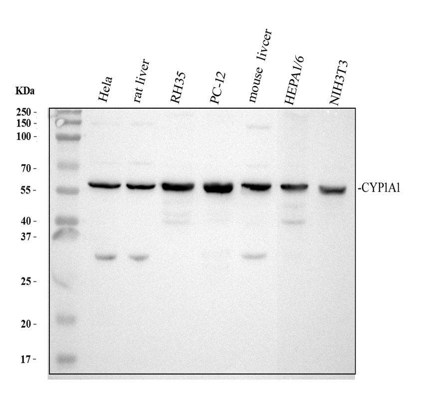
Western blot analysis of CYP1A1 using anti-CYP1A1 antibody. The sample well of each lane was loaded with 30 ug of sample under reducing conditions. Lane 1: human Hela whole cell lysates, Lane 2: rat liver tissue lysates, Lane 3: rat RH-35 whole cell lysates, Lane 4: rat PC-12 whole cell lysates, Lane 5: mouse liver tissue lysates, Lane 6: mouse HEPA1/6 whole cell lysates, Lane 7: mouse NIH/3T3 whole cell lysates. After electrophoresis, proteins were transferred to a membrane. Then the membrane was incubated with rabbit anti-CYP1A1 antigen affinity purified polyclonal antibody at a dilution of 1:1000 and probed with a goat anti-rabbit IgG-HRP secondary antibody. The signal is developed using ECL Plus Western Blotting Substrate . A specific band was detected for CYP1A1 at approximately 58 kDa. The expected band size for CYP1A1 is at 58 kDa.IHC
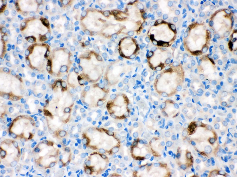
IHC analysis of CYP1A1 using anti-CYP1A1 antibody. CYP1A1 was detected in a paraffin-embedded section of rat kidney tissue. Biotinylated goat anti-rabbit IgG was used as secondary antibody. The tissue section was incubated with rabbit anti-CYP1A1 Antibody at a dilution of 1:200 and developed using Strepavidin-Biotin-Complex (SABC) with DAB as the chromogen.FC
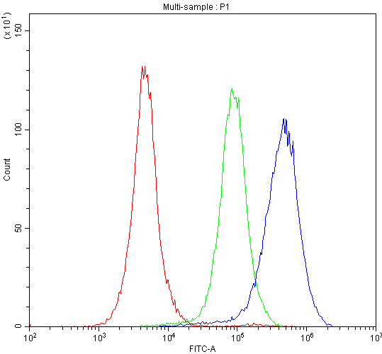
Flow Cytometry analysis of Caco-2 cells using anti-CYP1A1 antibody. Overlay histogram showing Caco-2 cells stained with PB9544 (Blue line). To facilitate intracellular staining, cells were fixed with 4% paraformaldehyde and permeabilized with permeabilization buffer. The cells were blocked with 10% normal goat serum. And then incubated with rabbit anti-CYP1A1 Antibody at 1:100 dilution for 30 min at 20°C. Fluoro488 conjugated goat anti-rabbit IgG was used as secondary antibody at 1:100 dilution for 30 minutes at 20°C. Isotype control antibody (Green line) was rabbit IgG at 1:100 dilution used under the same conditions. Unlabelled sample without incubation with primary antibody and secondary antibody (Red line) was used as a blank control.IF
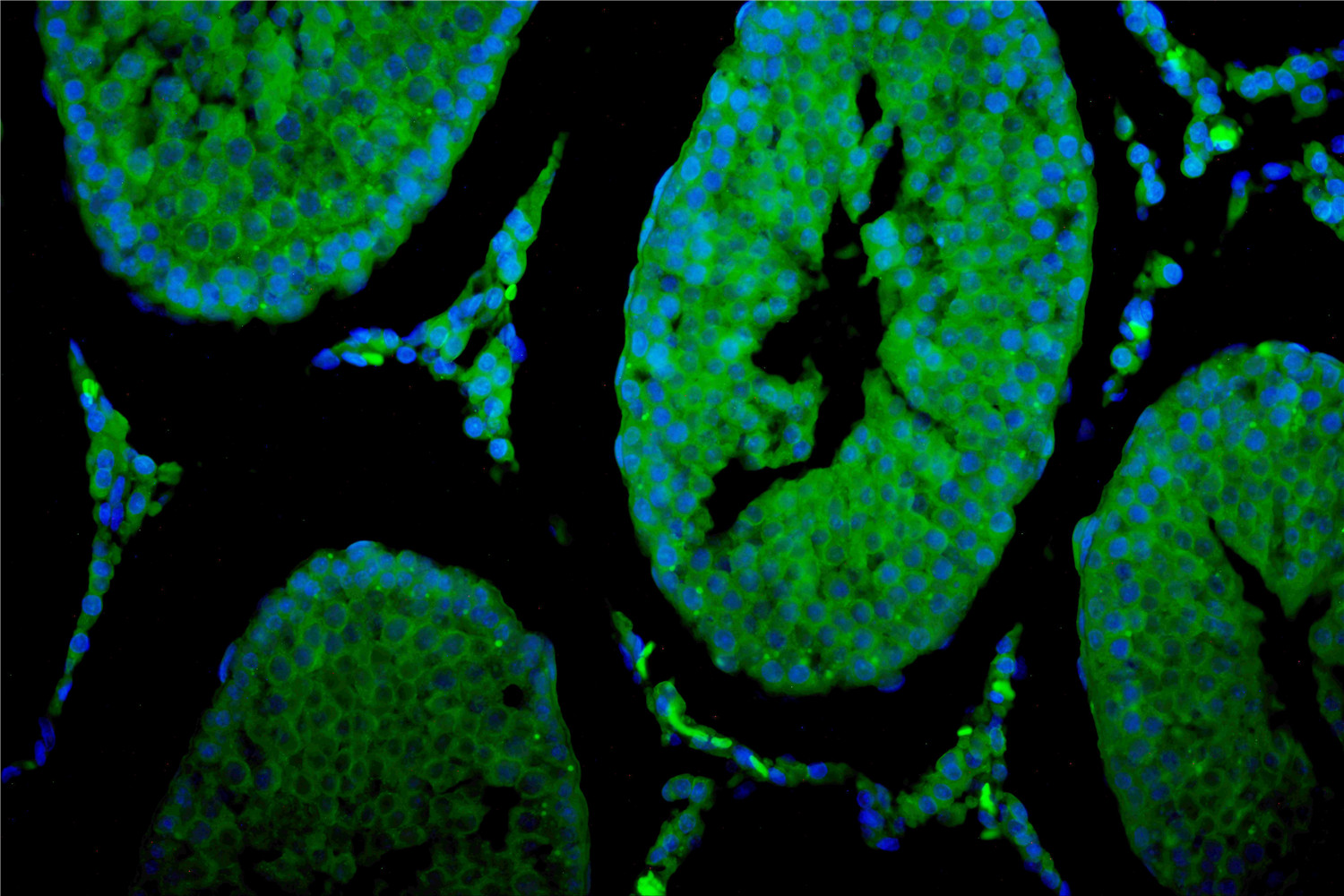
IF analysis using anti-CYP1A1 antibody . detected in paraffin-embedded section of rat testis tissue. The tissue section were stained using the Fluoro488 conjugated Anti-rabbit IgG Secondary Antibody (green)and counterstained with DAPI (blue).ICC/IF
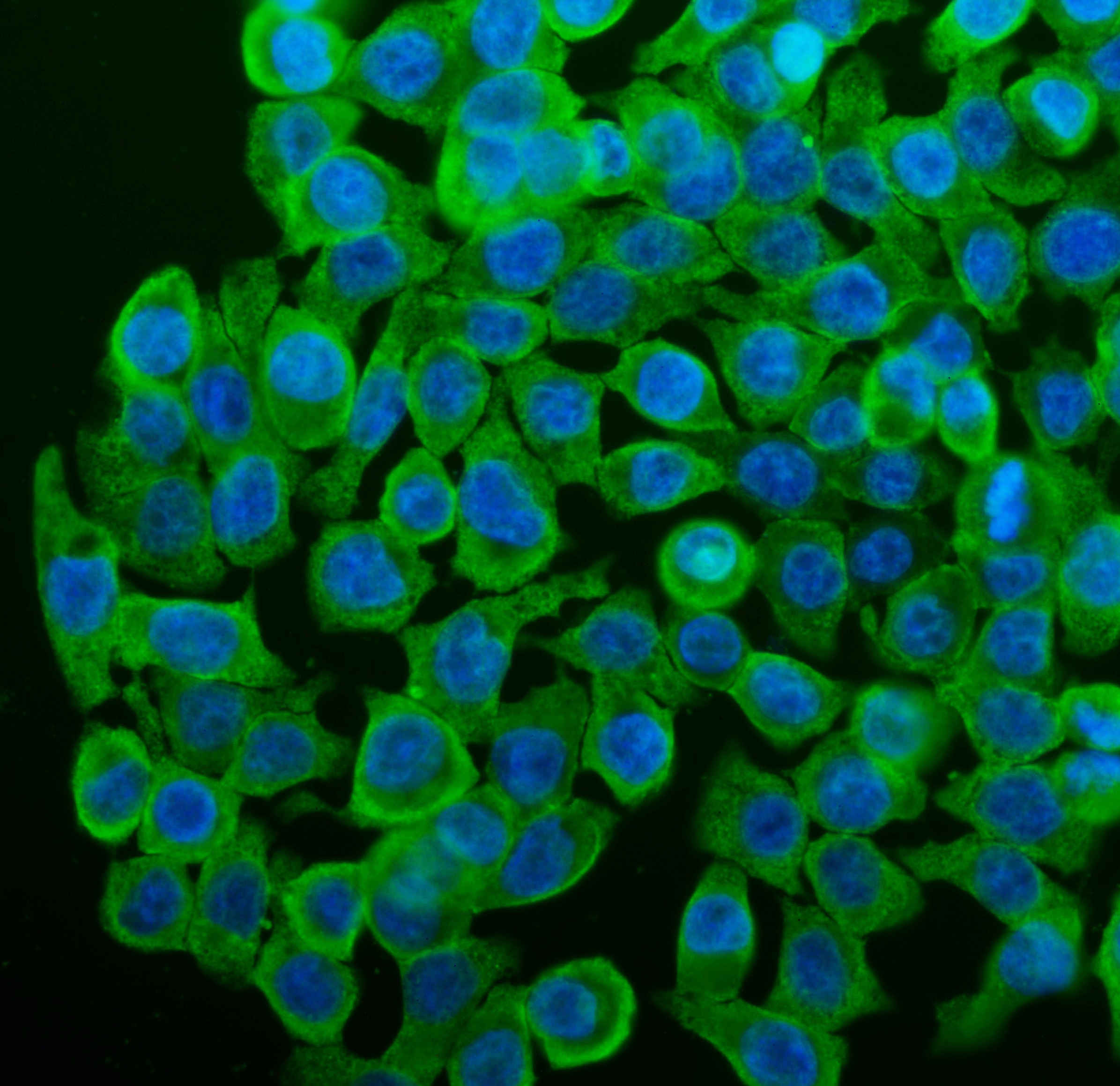
ICC/IF analysis of CYP1A1 using anti-CYP1A1 antibody. CYP1A1 was detected in an immunocytochemical section of Caco-2 cells. The section was incubated with rabbit anti-CYP1A1 Antibody at a dilution of 1:100. Fluoro488 Conjugated Goat Anti-Rabbit IgG (Green) was used as secondary antibody. The section was counterstained with DAPI(Blue).| Product Name | Anti-CYP1A1 Antibody |
|---|---|
| Antibody Type | Primary Antibodies |
| Immunogen | E.coli-derived human CYP1A1 recombinant protein (Position: H183-D320). Human CYP1A1 shares 81.2% amino acid (aa) sequence identity with both mouse and rat CYP1A1. |
| Clonality | Polyclonal |
|---|---|
| Isotype | IgG |
| Host Species | Rabbit |
| Tested Applications | FCICC/IFIFIHCWB |
| WB:1:500-2000 ICC/IF:1:50-1:400 FC:1:50-200 IHC:1:50-1:400 IF:1:50-1:400 |
|
| Species Reactivity | HumanMouseRat |
| Concentration | 0.5mg/ml |
| Purification | Affinity purified |
| Gene Symbol | CYP1A1 |
|---|---|
| Gene Synonyms | AHH CP11 CYP1 CYPIA1 P1-450 P450-C P450DX |
| Gene Full Name | cytochrome P450 family 1 subfamily A member 1 |
| Gene Summary | This gene, CYP1A1, encodes a member of the cytochrome P450 superfamily of enzymes. The cytochrome P450 proteins are monooxygenases which catalyze many reactions involved in drug metabolism and synthesis of cholesterol, steroids and other lipids. This protein localizes to the endoplasmic reticulum and its expression is induced by some polycyclic aromatic hydrocarbons (PAHs), some of which are found in cigarette smoke. The enzyme's endogenous substrate is unknown; however, it is able to metabolize some PAHs to carcinogenic intermediates. The gene has been associated with lung cancer risk. A related family member, CYP1A2, is located approximately 25 kb away from CYP1A1 on chromosome 15. Alternative splicing results in multiple transcript variants encoding distinct isoforms. [provided by RefSeq, Jan 2016] |
| Alternative Names | AHH AHRR CP11 CYP1 CYP1A1 CYPIA1 Cytochrome P450 1A1 Cytochrome P450 C Cytochrome P450 form 6 Cytochrome P450 P1 P1 450 P450 C P450DX |
| Molecular Weight(MW) | 58 kDa(Observed band size:57 kDa) |
| Cellular Localization | Cytoplasm,Endoplasmic reticulum,Membrane,Microsome,Mitochondrion,Mitochondrion inner membrane |
WB
Western blot analysis of CYP1A1 using anti-CYP1A1 antibody. The sample well of each lane was loaded with 30 ug of sample under reducing conditions. Lane 1: human Hela whole cell lysates, Lane 2: rat liver tissue lysates, Lane 3: rat RH-35 whole cell lysates, Lane 4: rat PC-12 whole cell lysates, Lane 5: mouse liver tissue lysates, Lane 6: mouse HEPA1/6 whole cell lysates, Lane 7: mouse NIH/3T3 whole cell lysates. After electrophoresis, proteins were transferred to a membrane. Then the membrane was incubated with rabbit anti-CYP1A1 antigen affinity purified polyclonal antibody at a dilution of 1:1000 and probed with a goat anti-rabbit IgG-HRP secondary antibody. The signal is developed using ECL Plus Western Blotting Substrate . A specific band was detected for CYP1A1 at approximately 58 kDa. The expected band size for CYP1A1 is at 58 kDa.
IHC
IHC analysis of CYP1A1 using anti-CYP1A1 antibody. CYP1A1 was detected in a paraffin-embedded section of rat kidney tissue. Biotinylated goat anti-rabbit IgG was used as secondary antibody. The tissue section was incubated with rabbit anti-CYP1A1 Antibody at a dilution of 1:200 and developed using Strepavidin-Biotin-Complex (SABC) with DAB as the chromogen.
FC
Flow Cytometry analysis of Caco-2 cells using anti-CYP1A1 antibody. Overlay histogram showing Caco-2 cells stained with PB9544 (Blue line). To facilitate intracellular staining, cells were fixed with 4% paraformaldehyde and permeabilized with permeabilization buffer. The cells were blocked with 10% normal goat serum. And then incubated with rabbit anti-CYP1A1 Antibody at 1:100 dilution for 30 min at 20°C. Fluoro488 conjugated goat anti-rabbit IgG was used as secondary antibody at 1:100 dilution for 30 minutes at 20°C. Isotype control antibody (Green line) was rabbit IgG at 1:100 dilution used under the same conditions. Unlabelled sample without incubation with primary antibody and secondary antibody (Red line) was used as a blank control.
IF
IF analysis using anti-CYP1A1 antibody . detected in paraffin-embedded section of rat testis tissue. The tissue section were stained using the Fluoro488 conjugated Anti-rabbit IgG Secondary Antibody (green)and counterstained with DAPI (blue).
ICC/IF
ICC/IF analysis of CYP1A1 using anti-CYP1A1 antibody. CYP1A1 was detected in an immunocytochemical section of Caco-2 cells. The section was incubated with rabbit anti-CYP1A1 Antibody at a dilution of 1:100. Fluoro488 Conjugated Goat Anti-Rabbit IgG (Green) was used as secondary antibody. The section was counterstained with DAPI(Blue).| Application Notes | WB:1:500-2000 ICC/IF:1:50-1:400 FC:1:50-200 IHC:1:50-1:400 IF:1:50-1:400 |
|---|
| Form | Liquid |
|---|---|
| Storage Instructions | 12 months from date of receipt,-20℃ as supplied. |
| Storage Buffer | 500 ug/ml antibody with PBS, 0.02% NaN3, 1 mg/ml BSA and 50% glycerol. |


