WB
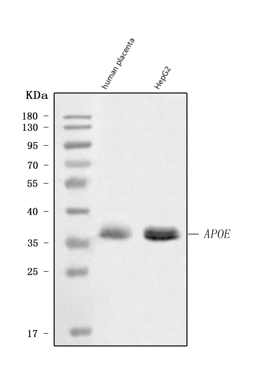
Western blot analysis of anti- APOE antibody. The sample well of each lane was loaded with 30ug of sample under reducing conditions. Lane 1: human placenta tissue lysates, Lane 2: human HepG2 whole cell lysates. Use rabbit anti- APOE 1:1000, probed with a goat anti-rabbit IgG-HRP secondary antibody. The signal is developed using an Enhanced Chemiluminescent detection (ECL) kit. A specific band was detected for APOE at approximately 36KD. The expected band size for APOE is at 36KD.IHC
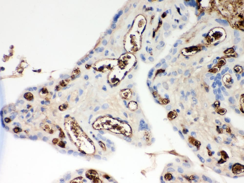
IHC analysis of APOE using anti-APOE antibody. APOE was detected in a paraffin-embedded section of human placenta tissue. Biotinylated goat anti-rabbit IgG was used as secondary antibody. The tissue section was incubated with rabbit anti-APOE Antibody at a dilution of 1:200 and developed using Strepavidin-Biotin-Complex (SABC) with DAB ( as the chromogen.IF
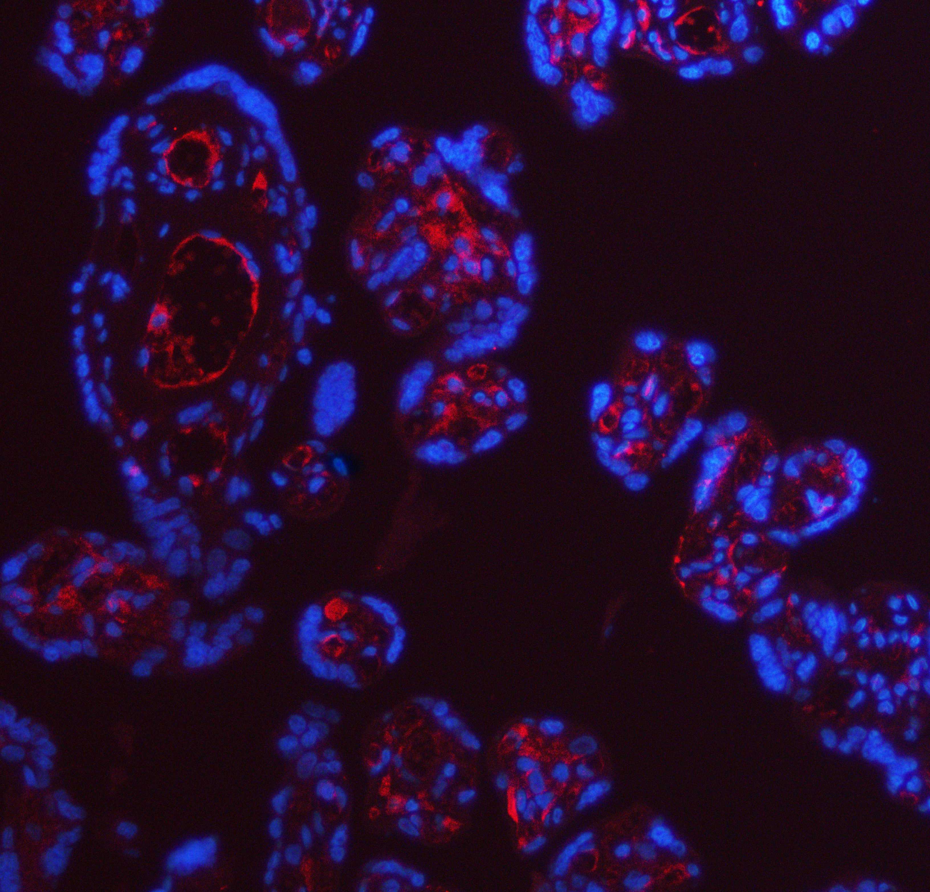
IF analysis of APOE using anti-APOE antibody. APOE was detected in a paraffin-embedded section of human placenta tissue. Cy3-conjugated Anti-rabbit IgG Secondary Antibody (red) was used as secondary antibody. The section was counterstained with DAPI (Blue).FC
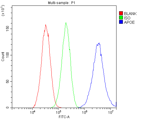
Flow Cytometry analysis of HepG2 cells using anti-APOE antibody. Overlay histogram showing HepG2 cells stained with (Blue line). To facilitate intracellular staining, cells were fixed with 4% paraformaldehyde and permeabilized with permeabilization buffer. The cells were blocked with 10% normal goat serum. And then incubated with rabbit anti-APOE Antibody at 1:100 dilution for 30 min at 20°C. Fluoro488 conjugated goat anti-rabbit IgG was used as secondary antibody at 1:100 dilution for 30 minutes at 20°C. Isotype control antibody (Green line) was rabbit IgG at 1:100 dilution used under the same conditions. Unlabelled sample without incubation with primary antibody and secondary antibody (Red line) was used as a blank control.ELISA
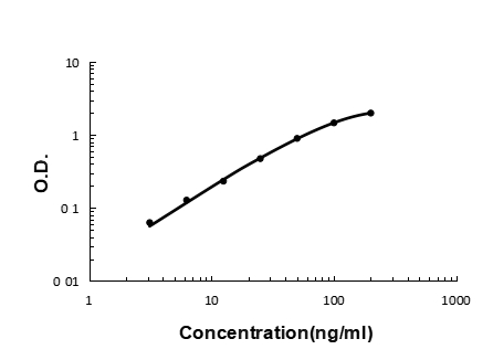
Sandwich ELISA - Recombinant human Apolipoprotein E/APOE protein standard curve. Use in combination with reagents from human APOE ELISA Kit EZ-Set (DIY Antibody Pairs).| Product Name | Anti-APOE Antibody |
|---|---|
| Antibody Type | Primary Antibodies |
| Immunogen | E.coli-derived human Apolipoprotein E recombinant protein (Position: K19-H317). Human Apolipoprotein E shares 73% and 72% amino acid (aa) sequence identity with mouse and rat Apolipoprotein E, respectively. |
| Clonality | Polyclonal |
|---|---|
| Isotype | IgG |
| Host Species | Rabbit |
| Tested Applications | ELISAFCIFIHCWB |
| WB:1:500-2000 FC:1:50-200 IHC:1:50-400 IF:1:50-400 ELISA:1:50-1:200 |
|
| Species Reactivity | Human |
| Concentration | 0.5mg/ml |
| Purification | Affinity purified |
| Gene Symbol | APOE |
|---|---|
| Gene Synonyms | AD2 LPG APO-E ApoE4 LDLCQ5 |
| Gene Full Name | apolipoprotein E |
| Gene Summary | The protein encoded by this gene is a major apoprotein of the chylomicron. It binds to a specific liver and peripheral cell receptor, and is essential for the normal catabolism of triglyceride-rich lipoprotein constituents. This gene maps to chromosome 19 in a cluster with the related apolipoprotein C1 and C2 genes. Mutations in this gene result in familial dysbetalipoproteinemia, or type III hyperlipoproteinemia (HLP III), in which increased plasma cholesterol and triglycerides are the consequence of impaired clearance of chylomicron and VLDL remnants. [provided by RefSeq, Jun 2016] |
| Alternative Names | AD2 Apo E APOE apoe4 apolipoprotein E LPG apolipoprotein E3 |
| Molecular Weight(MW) | 36 kDa |
| Cellular Localization | Chylomicron,Endosome,Extracellular matrix,HDL,Secreted,VLDL |
WB
Western blot analysis of anti- APOE antibody. The sample well of each lane was loaded with 30ug of sample under reducing conditions. Lane 1: human placenta tissue lysates, Lane 2: human HepG2 whole cell lysates. Use rabbit anti- APOE 1:1000, probed with a goat anti-rabbit IgG-HRP secondary antibody. The signal is developed using an Enhanced Chemiluminescent detection (ECL) kit. A specific band was detected for APOE at approximately 36KD. The expected band size for APOE is at 36KD.
IHC
IHC analysis of APOE using anti-APOE antibody. APOE was detected in a paraffin-embedded section of human placenta tissue. Biotinylated goat anti-rabbit IgG was used as secondary antibody. The tissue section was incubated with rabbit anti-APOE Antibody at a dilution of 1:200 and developed using Strepavidin-Biotin-Complex (SABC) with DAB ( as the chromogen.
IF
IF analysis of APOE using anti-APOE antibody. APOE was detected in a paraffin-embedded section of human placenta tissue. Cy3-conjugated Anti-rabbit IgG Secondary Antibody (red) was used as secondary antibody. The section was counterstained with DAPI (Blue).
FC
Flow Cytometry analysis of HepG2 cells using anti-APOE antibody. Overlay histogram showing HepG2 cells stained with (Blue line). To facilitate intracellular staining, cells were fixed with 4% paraformaldehyde and permeabilized with permeabilization buffer. The cells were blocked with 10% normal goat serum. And then incubated with rabbit anti-APOE Antibody at 1:100 dilution for 30 min at 20°C. Fluoro488 conjugated goat anti-rabbit IgG was used as secondary antibody at 1:100 dilution for 30 minutes at 20°C. Isotype control antibody (Green line) was rabbit IgG at 1:100 dilution used under the same conditions. Unlabelled sample without incubation with primary antibody and secondary antibody (Red line) was used as a blank control.
ELISA
Sandwich ELISA - Recombinant human Apolipoprotein E/APOE protein standard curve. Use in combination with reagents from human APOE ELISA Kit EZ-Set (DIY Antibody Pairs).| Application Notes | WB:1:500-2000 FC:1:50-200 IHC:1:50-400 IF:1:50-400 ELISA:1:50-1:200 |
|---|
| Form | Liquid |
|---|---|
| Storage Instructions | 12 months from date of receipt,-20℃ as supplied. |
| Storage Buffer | 500 ug/ml antibody with PBS, 0.02% NaN3, 1 mg/ml BSA and 50% glycerol. |


