WB
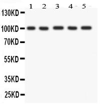
Western blot analysis of FGFR4 using anti-FGFR4 antibody. The sample well of each lane was loaded with 30 ug of sample under reducing conditions. Lane 1: HELA whole cell lysates, Lane 2: PANC whole cell lysates, Lane 3: SGC whole cell lysates, Lane 4: COLO320 whole cell lysates, Lane 5: SW620 whole cell lysates. After electrophoresis, proteins were transferred to a membrane. Then the membrane was incubated with rabbit anti-FGFR4 antigen affinity purified polyclonal antibody at a dilution of 1:1000 and probed with a goat anti-rabbit IgG-HRP secondary antibody. The signal is developed using ECL Plus Western Blotting Substrate. A specific band was detected for FGFR4 at approximately 100 kDa. The expected band size for FGFR4 is at 88 kDa.IHC
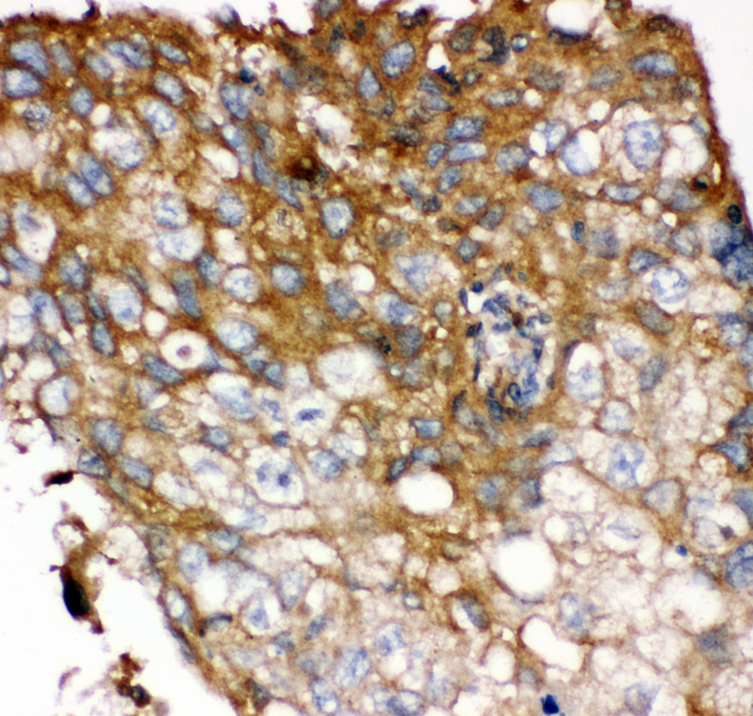
IHC analysis of FGFR4 using anti-FGFR4 antibody. FGFR4 was detected in a paraffin-embedded section of human lung cancer tissue. Biotinylated goat anti-rabbit IgG was used as secondary antibody. The tissue section was incubated with rabbit anti-FGFR4 Antibody at a dilution of 1:200 and developed using Strepavidin-Biotin-Complex (SABC) with DAB as the chromogen.| Product Name | Anti-FGFR4 Antibody |
|---|---|
| Antibody Type | Primary Antibodies |
| Immunogen | E.coli-derived human FGFR4 recombinant protein (Position: L22-H206). Human FGFR4 shares 86% and 84% amino acid (aa) sequences identity with mouse and rat FGFR4, respectively. |
| Clonality | Polyclonal |
|---|---|
| Isotype | IgG |
| Host Species | Rabbit |
| Tested Applications | IHCWB |
| WB:1:500-2000 IHC:1:50-400 |
|
| Species Reactivity | Human |
| Concentration | 0.5mg/ml |
| Purification | Affinity purified |
| Gene Symbol | FGFR4 |
|---|---|
| Gene Synonyms | TKF JTK2 CD334 |
| Gene Full Name | fibroblast growth factor receptor 4 |
| Gene Summary | The protein encoded by this gene is a tyrosine kinase and cell surface receptor for fibroblast growth factors. The encoded protein is involved in the regulation of several pathways, including cell proliferation, cell differentiation, cell migration, lipid metabolism, bile acid biosynthesis, vitamin D metabolism, glucose uptake, and phosphate homeostasis. This protein consists of an extracellular region, composed of three immunoglobulin-like domains, a single hydrophobic membrane-spanning segment, and a cytoplasmic tyrosine kinase domain. The extracellular portion interacts with fibroblast growth factors, setting in motion a cascade of downstream signals, ultimately influencing mitogenesis and differentiation. [provided by RefSeq, Aug 2017] |
| Alternative Names | CD334 FGFR 4 FGFR4 JTK2 TKF |
| Molecular Weight(MW) | 88 kDa(Observed band size:100 kDa) |
| Cellular Localization | Cell membrane,Endoplasmic reticulum,Endosome,Membrane,Secreted |
WB
Western blot analysis of FGFR4 using anti-FGFR4 antibody. The sample well of each lane was loaded with 30 ug of sample under reducing conditions. Lane 1: HELA whole cell lysates, Lane 2: PANC whole cell lysates, Lane 3: SGC whole cell lysates, Lane 4: COLO320 whole cell lysates, Lane 5: SW620 whole cell lysates. After electrophoresis, proteins were transferred to a membrane. Then the membrane was incubated with rabbit anti-FGFR4 antigen affinity purified polyclonal antibody at a dilution of 1:1000 and probed with a goat anti-rabbit IgG-HRP secondary antibody. The signal is developed using ECL Plus Western Blotting Substrate. A specific band was detected for FGFR4 at approximately 100 kDa. The expected band size for FGFR4 is at 88 kDa.
IHC
IHC analysis of FGFR4 using anti-FGFR4 antibody. FGFR4 was detected in a paraffin-embedded section of human lung cancer tissue. Biotinylated goat anti-rabbit IgG was used as secondary antibody. The tissue section was incubated with rabbit anti-FGFR4 Antibody at a dilution of 1:200 and developed using Strepavidin-Biotin-Complex (SABC) with DAB as the chromogen.| Application Notes | WB:1:500-2000 IHC:1:50-400 |
|---|
| Form | Liquid |
|---|---|
| Storage Instructions | 12 months from date of receipt,-20℃ as supplied. |
| Storage Buffer | 500 ug/ml antibody with PBS, 0.02% NaN3, 1 mg/ml BSA and 50% glycerol. |


