FC
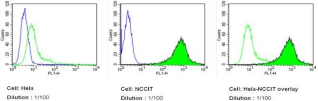
SSEA4 Antibody in Flow Flow cytometry analysis of SSEA-4 in NCCIT (green filled histogram) and HeLa cells (green unfilled histogram) compared to unstained cells (blue histogram). Positive staining is observed on NCCIT cells when compared to no antibody control (middle panel) and to SSEA-4-negative HeLa cells (right panel). Cells were fixed with 2% paraformaldehyde and washed with PBS. Cells were blocked with 2% BSA-PBS for 30 minutes at room temperature and incubated with a DyLight 488-conjugated SSEA-4 monoclonal antibody in 2% BSA-PBS at a dilution of 1:100 for 60 minutes at room temperature. Cells were washed and re-suspended in PBS for FACS analysis.SSEA4 Antibody in ICC/IF
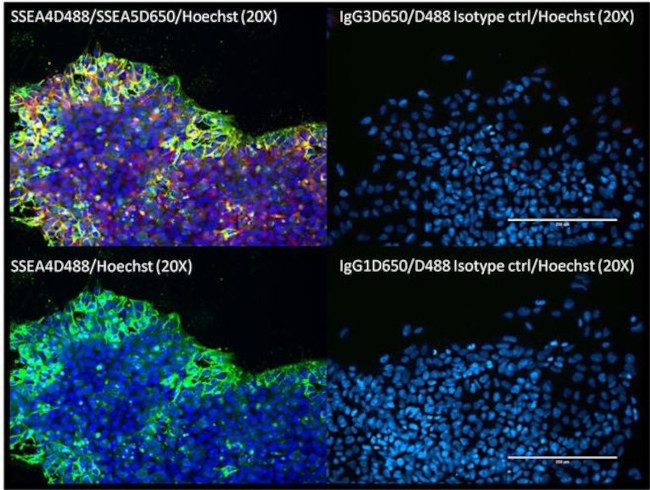
Imunofluorescent analysis of SSEA4 (green) in human episomal iPSC cells. Cells were grown on vitronectin coated plates, fixed with 4% paraformaldehyde for 15 minutes, and blocked with 3% Blocker BSA for 30 minutes at room temperature. Cells were stained with DyLight 488-conjugated SSEA4 monoclonal antibody (left panels) or DyLight 488-conjugated Mouse IgG3 isotype control antibody (top right panel), at a dilution of 1:50 and incubated for 1 hour at room temperature. Nuclei (blue) were stained with Hoechst 33342 dye. Images were taken on an EVOS FL Imaging system at 20X magnification.SSEA4 Antibody in ICC/IF
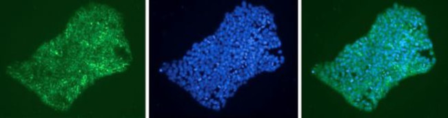
Immunofluorescent analysis of SSEA-4 (green) shows staining on the cell surface of human HEL 11.4 iPS cells, indicating pluripotency. SSEA-4 staining (left panel), DAPI staining (middle panel) and an overlay image of SSEA-4 with DAPI (right panel) are shown. HEL 11.4 iPS cells were grown on matrigel-coated chamber slides and fixed with 4% formaldehyde for 15 minutes prior to staining. Cells were probed with a DyLight 488-conjugated SSEA-4 monoclonal antibody in 0.1% Tween-PBS at a dilution of 1:500, and incubated overnight at 4C. Images were taken at 20X magnification.SSEA4 Antibody in IHC
Developmental cell 2018 - Figure 5 FACT Depletion Increases Efficiency of Reprogramming Human Fibroblasts (A) Reprogramming with human secondary fibroblasts carrying a doxycycline (DOX)-inducible OCT4/SOX2/KLF4/c-MYC (OSKM) cassette that were transfected with siRNAs against human SSRP1 or SUPTH16 before DOX induction. (B) Alkaline phosphatase (AP) staining of stem cell colonies 21 days after DOX treatment. Control experiment is scrambled siRNA. (C) Quantification of iPSC colonies (6 replicates) based on SSEA4 immunostaining for each knockdown condition. Paired Student's t test was used for statistical comparison, * p < 0.05, *** p < 0.001. Error bars represent SD. (D) Histological characterization of teratomas derived from grafting SSRP1 or SUPT16H depletion-derived iPSCs in mice. Teratomas reached 1.5 cm 2 in size after 51-70 days and showed tissues of all three germ layers (ectoderm, endoderm, mesoderm). Scale bars represent 50 mum. (E) Forced expression of Ascl1, Brn2, and Mytl1 in human fibroblasts (Normal Human Dermal Fibroblats = NHDF cells) and SSRP1 or SUPT16H depletion enhances reprogramming of fibroblasts to neurons. One-way ANOVA test was used for statistical comparison, ** p < 0.01. Error bars represent SEM. See also Figure S5 .| Product Name | Anti-SSEA4 Monoclonal Antibody Conjugated DL488 |
|---|---|
| Antibody Type | Primary Antibodies |
| Immunogen | human embryonal carcinoma cell line 2102Ep |
| Clonality | Monoclonal |
|---|---|
| Isotype | IgG3 |
| Host Species | Mouse |
| Tested Applications | FCICC/IFIHC |
| FC:1:50-1:500 ICC/IF:1:50-1:500 |
|
| Species Reactivity | Human |
| Concentration | 1mg/ml |
| Purification | Affinity purified |
| Gene Summary | SSEA-4 (Stage-specific embryonic antigen 4) is a cell surface antigen, expressed along with SSEA-3, TRA-1-60 and TRA-1-81 in embryonic stem cells, embryonal carcinoma cells and induced pluripotent stem cells (iPS). In human cells, these surface markers are down-regulated during the differentiation process, while SSEA-1 is absent in undifferentiated human stem cells and upregulated on the cell surface after retinoic acid mediated differentiation. Conversely, in mouse stem cells, SSEA-1 expression decreases with differentiation while SSEA-3 and SSEA-4 expression increase. |
|---|
FC
SSEA4 Antibody in Flow Flow cytometry analysis of SSEA-4 in NCCIT (green filled histogram) and HeLa cells (green unfilled histogram) compared to unstained cells (blue histogram). Positive staining is observed on NCCIT cells when compared to no antibody control (middle panel) and to SSEA-4-negative HeLa cells (right panel). Cells were fixed with 2% paraformaldehyde and washed with PBS. Cells were blocked with 2% BSA-PBS for 30 minutes at room temperature and incubated with a DyLight 488-conjugated SSEA-4 monoclonal antibody in 2% BSA-PBS at a dilution of 1:100 for 60 minutes at room temperature. Cells were washed and re-suspended in PBS for FACS analysis.
SSEA4 Antibody in ICC/IF
Imunofluorescent analysis of SSEA4 (green) in human episomal iPSC cells. Cells were grown on vitronectin coated plates, fixed with 4% paraformaldehyde for 15 minutes, and blocked with 3% Blocker BSA for 30 minutes at room temperature. Cells were stained with DyLight 488-conjugated SSEA4 monoclonal antibody (left panels) or DyLight 488-conjugated Mouse IgG3 isotype control antibody (top right panel), at a dilution of 1:50 and incubated for 1 hour at room temperature. Nuclei (blue) were stained with Hoechst 33342 dye. Images were taken on an EVOS FL Imaging system at 20X magnification.
SSEA4 Antibody in ICC/IF
Immunofluorescent analysis of SSEA-4 (green) shows staining on the cell surface of human HEL 11.4 iPS cells, indicating pluripotency. SSEA-4 staining (left panel), DAPI staining (middle panel) and an overlay image of SSEA-4 with DAPI (right panel) are shown. HEL 11.4 iPS cells were grown on matrigel-coated chamber slides and fixed with 4% formaldehyde for 15 minutes prior to staining. Cells were probed with a DyLight 488-conjugated SSEA-4 monoclonal antibody in 0.1% Tween-PBS at a dilution of 1:500, and incubated overnight at 4C. Images were taken at 20X magnification.
SSEA4 Antibody in IHC
Developmental cell 2018 - Figure 5 FACT Depletion Increases Efficiency of Reprogramming Human Fibroblasts (A) Reprogramming with human secondary fibroblasts carrying a doxycycline (DOX)-inducible OCT4/SOX2/KLF4/c-MYC (OSKM) cassette that were transfected with siRNAs against human SSRP1 or SUPTH16 before DOX induction. (B) Alkaline phosphatase (AP) staining of stem cell colonies 21 days after DOX treatment. Control experiment is scrambled siRNA. (C) Quantification of iPSC colonies (6 replicates) based on SSEA4 immunostaining for each knockdown condition. Paired Student's t test was used for statistical comparison, * p < 0.05, *** p < 0.001. Error bars represent SD. (D) Histological characterization of teratomas derived from grafting SSRP1 or SUPT16H depletion-derived iPSCs in mice. Teratomas reached 1.5 cm 2 in size after 51-70 days and showed tissues of all three germ layers (ectoderm, endoderm, mesoderm). Scale bars represent 50 mum. (E) Forced expression of Ascl1, Brn2, and Mytl1 in human fibroblasts (Normal Human Dermal Fibroblats = NHDF cells) and SSRP1 or SUPT16H depletion enhances reprogramming of fibroblasts to neurons. One-way ANOVA test was used for statistical comparison, ** p < 0.01. Error bars represent SEM. See also Figure S5 .| Application Notes | FC:1:50-1:500 ICC/IF:1:50-1:500 |
|---|
| Form | Liquid |
|---|---|
| Storage Instructions | 4°C, do not freeze |
| Storage Buffer | PBS with proprietary stabilizer |


