WB
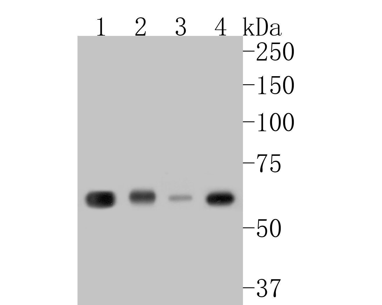
Western blot analysis of KLC1 on different lysates with Rabbit anti-KLC1 antibody at 1/500 dilution. Lane 1: Human brain tissue lysate (20 µg/Lane), Lane 2: Rat brain tissue lysate (20 µg/Lane), Lane 3: Hela cell lysate (10 µg/Lane), Lane 4: PC-12 cell lysate (10 µg/Lane), Exposure time: 2 minutes; 10% SDS-PAGE gel. Proteins were transferred to a PVDF membrane and blocked with 5% NFDM/TBST for 1 hour at room temperature. The primary antibody at 1/500 dilution was used in 5% NFDM/TBST at room temperature for 2 hours. Goat Anti-Rabbit IgG - HRP Secondary Antibody at 1/5000 dilution was used for 1 hour at room temperature.ICC/IF
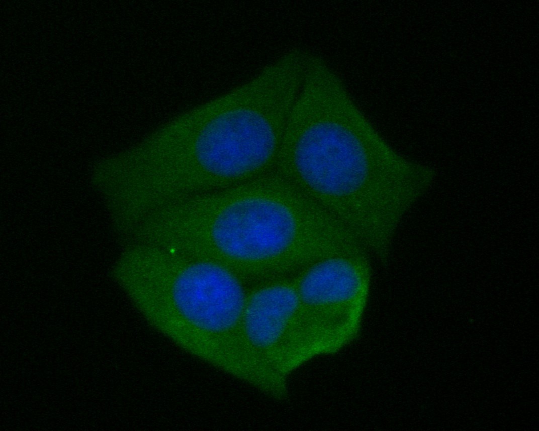
Immunocytochemistry analysis of MCF-7 cells labeling KLC1 with Rabbit anti-KLC1 antibody at 1/50 dilution. Cells were fixed in 4% paraformaldehyde for 10 minutes at 37 ℃, permeabilized with 0.05% Triton X-100 in PBS for 20 minutes, and then blocked with 2% negative goat serum for 30 minutes at room temperature. Cells were then incubated with Rabbit anti-KLC1 antibody at 1/50 dilution in 2% negative goat serum overnight at 4 ℃. 488 conjugate-Goat anti-Rabbit IgG was used as the secondary antibody at 1/1,000 dilution.Nuclear DNA was labelled in blue with DAPI.ICC/IF
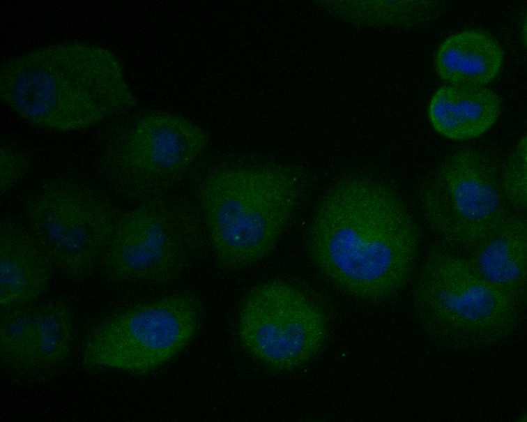
Immunocytochemistry analysis of RWPE-1 cells labeling KLC1 with Rabbit anti-KLC1 antibody at 1/50 dilution. Cells were fixed in 4% paraformaldehyde for 10 minutes at 37 ℃, permeabilized with 0.05% Triton X-100 in PBS for 20 minutes, and then blocked with 2% negative goat serum for 30 minutes at room temperature. Cells were then incubated with Rabbit anti-KLC1 antibody at 1/50 dilution in 2% negative goat serum overnight at 4 ℃.488 conjugate-Goat anti-Rabbit IgG was used as the secondary antibody at 1/1,000 dilution.Nuclear DNA was labelled in blue with DAPI.IHC
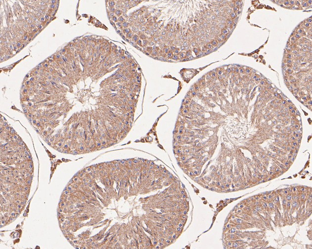
Immunohistochemical analysis of paraffin-embedded rat testis tissue with Rabbit anti-KLC1 antibody at 1/50 dilution. The section was pre-treated using heat mediated antigen retrieval with Tris-EDTA buffer (pH 8.0-8.4) for 20 minutes. The tissues were blocked in 1% BSA for 20 minutes at room temperature, washed with ddH2O and PBS, and then probed with the primary antibody at 1/50 dilution for 0.5 hour at room temperature. The detection was performed using an HRP conjugated compact polymer system. DAB was used as the chromogen. Tissues were counterstained with hematoxylin and mounted with DPX.IHC
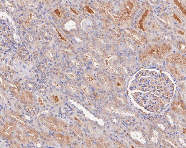
Immunohistochemical analysis of paraffin-embedded human kidney tissue with Rabbit anti-KLC1 antibody at 1/200 dilution. The section was pre-treated using heat mediated antigen retrieval with Tris-EDTA buffer (pH 8.0-8.4) for 20 minutes. The tissues were blocked in 1% BSA for 20 minutes at room temperature, washed with ddH2O and PBS, and then probed with the primary antibody at 1/200 dilution for 0.5 hour at room temperature. The detection was performed using an HRP conjugated compact polymer system. DAB was used as the chromogen. Tissues were counterstained with hematoxylin and mounted with DPX.IHC
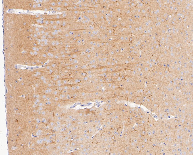
Immunohistochemical analysis of paraffin-embedded mouse brain tissue with Rabbit anti-KLC1 antibody at 1/400 dilution. The section was pre-treated using heat mediated antigen retrieval with Tris-EDTA buffer (pH 8.0-8.4) for 20 minutes. The tissues were blocked in 1% BSA for 20 minutes at room temperature, washed with ddH2O and PBS, and then probed with the primary antibody at 1/400 dilution for 0.5 hour at room temperature. The detection was performed using an HRP conjugated compact polymer system. DAB was used as the chromogen. Tissues were counterstained with hematoxylin and mounted with DPX.FC
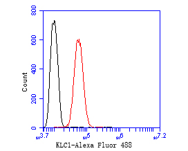
Flow cytometric analysis of KLC1 was done on SH-SY5Y cells. The cells were fixed, permeabilized and stained with the primary antibody (1/50) (red). After incubation of the primary antibody at room temperature for an hour, the cells were stained with a 488-conjugated Goat anti-Rabbit IgG Secondary antibody at 1/1000 dilution for 30 minutes.Unlabelled sample was used as a control (cells without incubation with primary antibody; black).| Product Name | KLC1 Recombinant Rabbit Monoclonal Antibody |
|---|---|
| Antibody Type | Primary Antibodies |
| Immunogen | Recombinant protein within Human KLC1 aa 461-573 / 573. |
| Clonality | monoclonal |
|---|---|
| Isotype | IgG |
| Host Species | Rabbit |
| Tested Applications | FCICC/IFIHCWB |
| WB:1:500-1:2000 ICC/IF:1:50-1:100 IHC:1:50-1:500 FC:1:50-1:100 |
|
| Species Reactivity | HumanMouseRat |
| Concentration | 1mg/ml |
| Purification | Protein A |
| Gene Symbol | KLC1 |
|---|---|
| Gene Synonyms | KLC KNS2 KNS2A |
| Gene Full Name | kinesin light chain 1 |
| Gene Summary | Conventional kinesin is a tetrameric molecule composed of two heavy chains and two light chains, and transports various cargos along microtubules toward their plus ends. The heavy chains provide the motor activity, while the light chains bind to various cargos. This gene encodes a member of the kinesin light chain family. It associates with kinesin heavy chain through an N-terminal domain, and six tetratricopeptide repeat (TPR) motifs are thought to be involved in binding of cargos such as vesicles, mitochondria, and the Golgi complex. Thus, kinesin light chains function as adapter molecules and not motors per se. Although previously named "kinesin 2", this gene is not a member of the kinesin-2 / kinesin heavy chain subfamily of kinesin motor proteins. Extensive alternative splicing produces isoforms with different C-termini that are proposed to bind to different cargos; however, the full-length nature and/or biological validity of most of these variants have not been determined. [provided by RefSeq, Jul 2008] |
| Alternative Names | Kinesin 2 60/70kDa antibody Kinesin light chain 1 antibody Kinesin2 antibody KLC 1 antibody KLC antibody Klc1 antibody KLC1_HUMAN antibody KNS 2 antibody KNS 2A antibody KNS2 antibody Kinesin 2 60/70kDa antibody Kinesin light chain 1 antibody Kinesin2 antibody KLC 1 antibody KLC antibody Klc1 antibody KLC1_HUMAN antibody KNS 2 antibody KNS 2A antibody KNS2 antibody KNS2A antibody Medulloblastoma antigen MU MB 2.50 antibody MGC15245 antibody |
| Molecular Weight(MW) | 65kDa(Observed band size: 65kDa) |
| Cellular Localization | Cytoskeleton, growth cone, cytoplasmic vesicle. |
WB
Western blot analysis of KLC1 on different lysates with Rabbit anti-KLC1 antibody at 1/500 dilution. Lane 1: Human brain tissue lysate (20 µg/Lane), Lane 2: Rat brain tissue lysate (20 µg/Lane), Lane 3: Hela cell lysate (10 µg/Lane), Lane 4: PC-12 cell lysate (10 µg/Lane), Exposure time: 2 minutes; 10% SDS-PAGE gel. Proteins were transferred to a PVDF membrane and blocked with 5% NFDM/TBST for 1 hour at room temperature. The primary antibody at 1/500 dilution was used in 5% NFDM/TBST at room temperature for 2 hours. Goat Anti-Rabbit IgG - HRP Secondary Antibody at 1/5000 dilution was used for 1 hour at room temperature.
ICC/IF
Immunocytochemistry analysis of MCF-7 cells labeling KLC1 with Rabbit anti-KLC1 antibody at 1/50 dilution. Cells were fixed in 4% paraformaldehyde for 10 minutes at 37 ℃, permeabilized with 0.05% Triton X-100 in PBS for 20 minutes, and then blocked with 2% negative goat serum for 30 minutes at room temperature. Cells were then incubated with Rabbit anti-KLC1 antibody at 1/50 dilution in 2% negative goat serum overnight at 4 ℃. 488 conjugate-Goat anti-Rabbit IgG was used as the secondary antibody at 1/1,000 dilution.Nuclear DNA was labelled in blue with DAPI.
ICC/IF
Immunocytochemistry analysis of RWPE-1 cells labeling KLC1 with Rabbit anti-KLC1 antibody at 1/50 dilution. Cells were fixed in 4% paraformaldehyde for 10 minutes at 37 ℃, permeabilized with 0.05% Triton X-100 in PBS for 20 minutes, and then blocked with 2% negative goat serum for 30 minutes at room temperature. Cells were then incubated with Rabbit anti-KLC1 antibody at 1/50 dilution in 2% negative goat serum overnight at 4 ℃.488 conjugate-Goat anti-Rabbit IgG was used as the secondary antibody at 1/1,000 dilution.Nuclear DNA was labelled in blue with DAPI.
IHC
Immunohistochemical analysis of paraffin-embedded rat testis tissue with Rabbit anti-KLC1 antibody at 1/50 dilution. The section was pre-treated using heat mediated antigen retrieval with Tris-EDTA buffer (pH 8.0-8.4) for 20 minutes. The tissues were blocked in 1% BSA for 20 minutes at room temperature, washed with ddH2O and PBS, and then probed with the primary antibody at 1/50 dilution for 0.5 hour at room temperature. The detection was performed using an HRP conjugated compact polymer system. DAB was used as the chromogen. Tissues were counterstained with hematoxylin and mounted with DPX.
IHC
Immunohistochemical analysis of paraffin-embedded human kidney tissue with Rabbit anti-KLC1 antibody at 1/200 dilution. The section was pre-treated using heat mediated antigen retrieval with Tris-EDTA buffer (pH 8.0-8.4) for 20 minutes. The tissues were blocked in 1% BSA for 20 minutes at room temperature, washed with ddH2O and PBS, and then probed with the primary antibody at 1/200 dilution for 0.5 hour at room temperature. The detection was performed using an HRP conjugated compact polymer system. DAB was used as the chromogen. Tissues were counterstained with hematoxylin and mounted with DPX.
IHC
Immunohistochemical analysis of paraffin-embedded mouse brain tissue with Rabbit anti-KLC1 antibody at 1/400 dilution. The section was pre-treated using heat mediated antigen retrieval with Tris-EDTA buffer (pH 8.0-8.4) for 20 minutes. The tissues were blocked in 1% BSA for 20 minutes at room temperature, washed with ddH2O and PBS, and then probed with the primary antibody at 1/400 dilution for 0.5 hour at room temperature. The detection was performed using an HRP conjugated compact polymer system. DAB was used as the chromogen. Tissues were counterstained with hematoxylin and mounted with DPX.
FC
Flow cytometric analysis of KLC1 was done on SH-SY5Y cells. The cells were fixed, permeabilized and stained with the primary antibody (1/50) (red). After incubation of the primary antibody at room temperature for an hour, the cells were stained with a 488-conjugated Goat anti-Rabbit IgG Secondary antibody at 1/1000 dilution for 30 minutes.Unlabelled sample was used as a control (cells without incubation with primary antibody; black).| Application Notes | WB:1:500-1:2000 ICC/IF:1:50-1:100 IHC:1:50-1:500 FC:1:50-1:100 |
|---|
| Form | Liquid |
|---|---|
| Storage Instructions | Shipped at 4℃. Store at +4℃ short term (1-2 weeks). It is recommended to aliquot into single-use upon delivery. Store at -20℃ long term. |
| Storage Buffer | 1*TBS (pH7.4), 0.05% BSA, 40% Glycerol. Preservative: 0.05% Sodium Azide. |


