WB
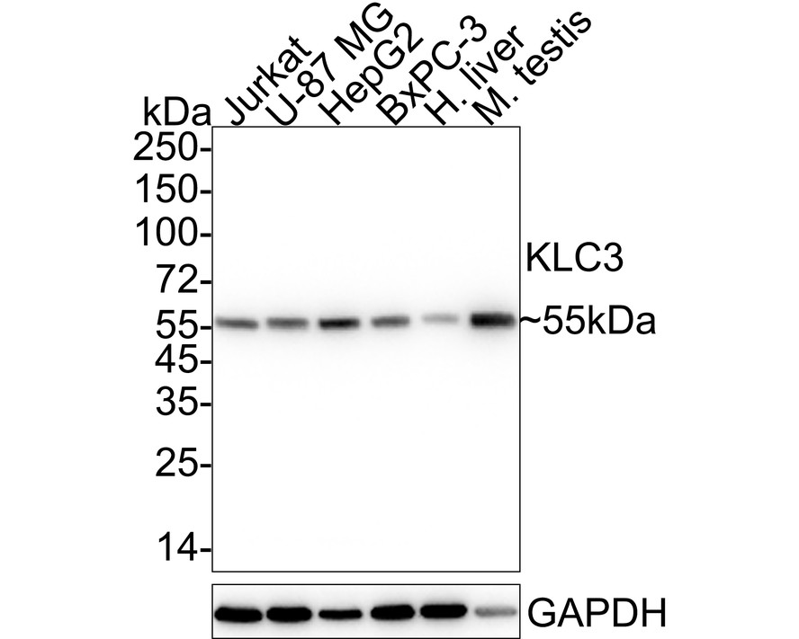
Western blot analysis of KLC3 on different lysates with Rabbit anti-KLC3 antibody at 1/1000 dilution. Lane 1: Jurkat cell lysate (20 µg/Lane), Lane 2: U-87 MG cell lysate (20 µg/Lane), Lane 3: HepG2 cell lysate (20 µg/Lane), Lane 4: BxPC-3 cell lysate (20 µg/Lane), Lane 5: Human liver tissue lysate (40 µg/Lane), Lane 6: Mouse testis tissue lysate (40 µg/Lane), Exposure time: 2 seconds; 4-20% SDS-PAGE gel. Proteins were transferred to a PVDF membrane and blocked with 5% NFDM/TBST for 1 hour at room temperature. The primary antibody at 1/1,000 dilution was used in 5% NFDM/TBST at 4℃ overnight. Goat Anti-Rabbit IgG - HRP Secondary Antibody at 1/50000 dilution was used for 1 hour at room temperature.ICC/IF
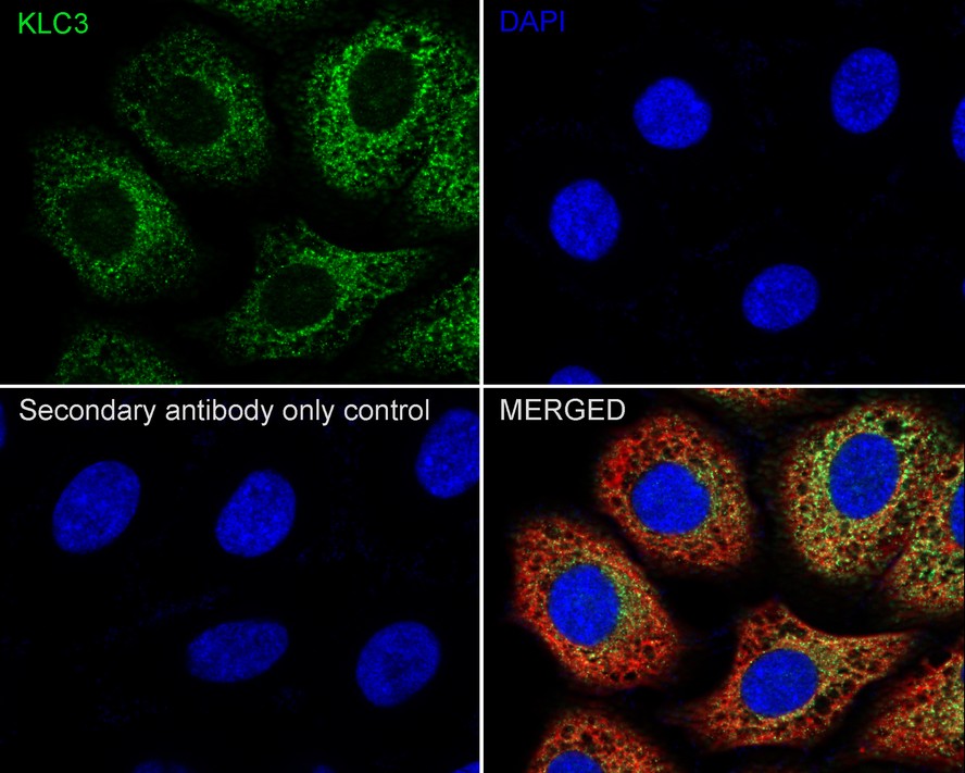
Immunocytochemistry analysis of HepG2 cells labeling KLC3 with Rabbit anti-KLC3 antibody at 1/500 dilution. Cells were fixed in 4% paraformaldehyde for 20 minutes at room temperature, permeabilized with 0.1% Triton X-100 in PBS for 5 minutes at room temperature, then blocked with 1% BSA in 10% negative goat serum for 1 hour at room temperature. Cells were then incubated with Rabbit anti-KLC3 antibody at 1/500 dilution in 1% BSA in PBST overnight at 4 ℃. Goat Anti-Rabbit IgG H&L (488) was used as the secondary antibody at 1/1,000 dilution. PBS instead of the primary antibody was used as the secondary antibody only control. Nuclear DNA was labelled in blue with DAPI. Beta tubulin (red) was stained at 1/100 dilution overnight at +4℃. Goat Anti-Mouse IgG H&L (594) was used as the secondary antibody at 1/1,000 dilution.FC
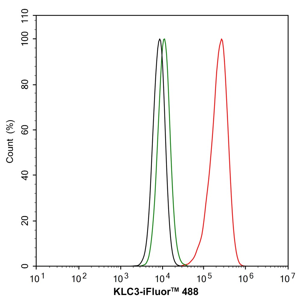
Flow cytometric analysis of HepG2 cells labeling KLC3. Cells were fixed and permeabilized. Then stained with the primary antibody (1/1000) (red) compared with Rabbit IgG Isotype Control (green). After incubation of the primary antibody at +4℃ for an hour, the cells were stained with a iFluor™ 488 conjugate-Goat anti-Rabbit IgG Secondary antibody at 1/1,000 dilution for 30 minutes at +4℃. Unlabelled sample was used as a control (cells without incubation with primary antibody; black).| Product Name | KLC3 Recombinant Rabbit Monoclonal Antibody |
|---|---|
| Antibody Type | Primary Antibodies |
| Immunogen | Synthetic peptide within Human KLC3 aa 350-450. |
| Clonality | monoclonal |
|---|---|
| Isotype | IgG |
| Host Species | Rabbit |
| Tested Applications | FCICC/IFWB |
| WB:1:1000 ICC/IF:1:500 FC:1:1000 |
|
| Species Reactivity | HumanMouseRat |
| Concentration | 1mg/ml |
| Purification | Protein A |
| Gene Symbol | KLC3 |
|---|---|
| Gene Synonyms | KLC2 KLCt KLC2L KNS2B |
| Gene Full Name | kinesin light chain 3 |
| Gene Summary | This gene encodes a member of the kinesin light chain gene family. Kinesins are molecular motors involved in the transport of cargo along microtubules, and are composed of two kinesin heavy chain (KHC) and two kinesin light chain (KLC) molecules. KLCs are thought to typically be involved in binding cargo and regulating kinesin activity. In the rat, a protein similar to this gene product is expressed in post-meiotic spermatids, where it associates with structural components of sperm tails and mitochondria. [provided by RefSeq, Jul 2008] |
| Alternative Names | Kinesin light chain 2 antibody Kinesin light chain 3 antibody KLC2 antibody KLC2 like antibody KLC2-like antibody KLC2L antibody KLC3 antibody KLC3_HUMAN antibody KLCt antibody KNS2B antibody |
| Molecular Weight(MW) | 55kDa(Observed band size: 55kDa) |
| Cellular Localization | Cytoplasm, cytoskeleton, Mitochondrion. |
WB
Western blot analysis of KLC3 on different lysates with Rabbit anti-KLC3 antibody at 1/1000 dilution. Lane 1: Jurkat cell lysate (20 µg/Lane), Lane 2: U-87 MG cell lysate (20 µg/Lane), Lane 3: HepG2 cell lysate (20 µg/Lane), Lane 4: BxPC-3 cell lysate (20 µg/Lane), Lane 5: Human liver tissue lysate (40 µg/Lane), Lane 6: Mouse testis tissue lysate (40 µg/Lane), Exposure time: 2 seconds; 4-20% SDS-PAGE gel. Proteins were transferred to a PVDF membrane and blocked with 5% NFDM/TBST for 1 hour at room temperature. The primary antibody at 1/1,000 dilution was used in 5% NFDM/TBST at 4℃ overnight. Goat Anti-Rabbit IgG - HRP Secondary Antibody at 1/50000 dilution was used for 1 hour at room temperature.
ICC/IF
Immunocytochemistry analysis of HepG2 cells labeling KLC3 with Rabbit anti-KLC3 antibody at 1/500 dilution. Cells were fixed in 4% paraformaldehyde for 20 minutes at room temperature, permeabilized with 0.1% Triton X-100 in PBS for 5 minutes at room temperature, then blocked with 1% BSA in 10% negative goat serum for 1 hour at room temperature. Cells were then incubated with Rabbit anti-KLC3 antibody at 1/500 dilution in 1% BSA in PBST overnight at 4 ℃. Goat Anti-Rabbit IgG H&L (488) was used as the secondary antibody at 1/1,000 dilution. PBS instead of the primary antibody was used as the secondary antibody only control. Nuclear DNA was labelled in blue with DAPI. Beta tubulin (red) was stained at 1/100 dilution overnight at +4℃. Goat Anti-Mouse IgG H&L (594) was used as the secondary antibody at 1/1,000 dilution.
FC
Flow cytometric analysis of HepG2 cells labeling KLC3. Cells were fixed and permeabilized. Then stained with the primary antibody (1/1000) (red) compared with Rabbit IgG Isotype Control (green). After incubation of the primary antibody at +4℃ for an hour, the cells were stained with a iFluor™ 488 conjugate-Goat anti-Rabbit IgG Secondary antibody at 1/1,000 dilution for 30 minutes at +4℃. Unlabelled sample was used as a control (cells without incubation with primary antibody; black).| Application Notes | WB:1:1000 ICC/IF:1:500 FC:1:1000 |
|---|
| Form | Liquid |
|---|---|
| Storage Instructions | Shipped at 4℃. Store at +4℃ short term (1-2 weeks). It is recommended to aliquot into single-use upon delivery. Store at -20℃ long term. |
| Storage Buffer | 1*TBS (pH7.4), 0.05% BSA, 40% Glycerol. Preservative: 0.05% Sodium Azide. |


